Accéder au contenu
Merck
Toutes les photos(1)

Key Documents

06-1334

Sigma-Aldrich

Anti-phospho-TAB1 (Thr431) Antibody

from rabbit, purified by affinity chromatography

Synonyme(s) :

TGF-beta activated kinase 1/MAP3K7 binding protein 1, TAK1-binding protein 1, transforming growth factor beta-activated kinase-binding protein 1, mitogen-activated protein kinase kinase kinase 7 interacting protein 1, Mitogen-activated protein kinase kin

Se connecterpour consulter vos tarifs contractuels et ceux de votre entreprise/organisme


About This Item

Code UNSPSC :
12352203
eCl@ss :
32160702
Nomenclature NACRES :
NA.41

Source biologique

rabbit

Niveau de qualité

Forme d'anticorps

affinity isolated antibody

Type de produit anticorps

primary antibodies

Clone

polyclonal

Produit purifié par

affinity chromatography

Espèces réactives

rat, human, horse, mouse

Réactivité de l'espèce (prédite par homologie)

equine (based on 100% sequence homology), ox (based on 100% sequence homology)

Technique(s)

western blot: suitable

Numéro d'accès NCBI

Numéro d'accès UniProt

Conditions d'expédition

wet ice

Modification post-traductionnelle de la cible

phosphorylation (pThr431)

Informations sur le gène

human ... TAB1(10454)

Description générale

TAK1 binding protein 1 (TAB1) is thought to activate TAK1 MAPKKK in transforming growth factor-beta (TGF-beta) signaling. TAB1 can also associate with TAK1 to form a unique MAPKKK complex that is thought to activate AP-1 and the NFkappaB signaling pathways. Through conformational changes, TAB1 is also thought to induce p38alpha autophosphorylation. It has been linked to both IL-1alpha and TNFalpha signaling and therefore may also be involved in the inflammatory response. Recent research has found that the TAK1-TAB1 complex may have anti-inflammatory effects by aiding stercurensin in regulating the NFκB-dependent inflammatory pathways.

Spécificité

This antibody recognizes TAB1 phosphorylated at Thr431.

Immunogène

Epitope: Phosphorylated Thr431
KLH-conjugated linear peptide corresponding to human TAB1 phosphorylated at Thr431.

Application

Research Category
Inflammation & Immunology
Research Sub Category
Immunoglobulins & Immunology
This Anti-phospho-TAB1 (Thr431) Antibody is validated for use in WB for the detection of phospho-TAB1 (Thr431).

Qualité

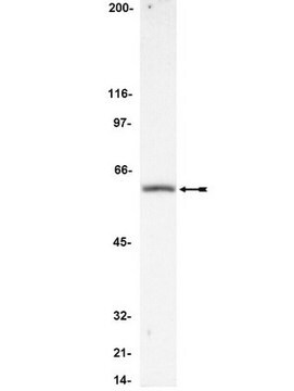
Evaluated by Western Blot in L6 cell lysate.

Western Blot Analysis: 1 µg/mL of this antibody detected TAB1 on 10 µg of L6 cell lysate.

Description de la cible

~78 kDa observed. 55 kDa calculated. A higher molecular weight on western blot may be due to glycosylation. An uncharacterized band appears at ~42 and 50 kDa in some lysates.

Forme physique

Affinity purified
Purified rabbit polyclonal in buffer containing 0.1 M Tris-Glycine (pH 7.4), 150 mM NaCl with 0.05% sodium azide.

Stockage et stabilité

Stable for 1 year at 2-8°C from date of receipt.

Remarque sur l'analyse

Control
L6 cell lysate

Autres remarques

Concentration: Please refer to the Certificate of Analysis for the lot-specific concentration.

Clause de non-responsabilité

Unless otherwise stated in our catalog or other company documentation accompanying the product(s), our products are intended for research use only and are not to be used for any other purpose, which includes but is not limited to, unauthorized commercial uses, in vitro diagnostic uses, ex vivo or in vivo therapeutic uses or any type of consumption or application to humans or animals.

Vous ne trouvez pas le bon produit ?  

Essayez notre Outil de sélection de produits.

Code de la classe de stockage

12 - Non Combustible Liquids

Classe de danger pour l'eau (WGK)

WGK 1

Point d'éclair (°F)

Not applicable

Point d'éclair (°C)

Not applicable


Certificats d'analyse (COA)

Recherchez un Certificats d'analyse (COA) en saisissant le numéro de lot du produit. Les numéros de lot figurent sur l'étiquette du produit après les mots "Lot" ou "Batch".

Déjà en possession de ce produit ?

Retrouvez la documentation relative aux produits que vous avez récemment achetés dans la Bibliothèque de documents.

Consulter la Bibliothèque de documents

Briana Ortiz et al.
American journal of reproductive immunology (New York, N.Y. : 1989), 89(3), e13664-e13664 (2022-12-11)
During gestation, the decidua is an essential layer of the maternal-fetal interface, providing immune support and maintaining inflammatory homeostasis. Although Chlamydia (C.) trachomatis is associated with adverse pregnancy outcomes the pathogenic effects on maternal decidua contributing to adverse events are
Bjarte Bergstrøm et al.
Journal of immunology (Baltimore, Md. : 1950), 195(3), 1100-1111 (2015-06-19)
Staphylococcus aureus may cause serious infections and is one of the most lethal and common causes of sepsis. TLR2 has been described as the main pattern recognition receptor that senses S. aureus and elicits production of proinflammatory cytokines via MyD88
Chia-Yu Yang et al.
Journal of immunology (Baltimore, Md. : 1950), 192(4), 1547-1557 (2014-01-10)
T cell activation is dependent upon phosphorylation of MAPKs, which play a critical role in the regulation of immune responses. Dual-specificity phosphatase 14 (DUSP14; also known as MKP6) is classified as a MAPK phosphatase. The in vivo functions of DUSP14
Mariana de Castro Silva et al.
Reproduction (Cambridge, England), 160(4), 627-638 (2020-08-26)
A non-reversible state of epithelial to mesenchymal transition (EMT) at term accumulates proinflammatory mesenchymal cells and predisposes fetal membrane to weakening prior to delivery at term. We investigated the induction of EMT in amnion epithelial cells (AEC) in response to

Notre équipe de scientifiques dispose d'une expérience dans tous les secteurs de la recherche, notamment en sciences de la vie, science des matériaux, synthèse chimique, chromatographie, analyse et dans de nombreux autres domaines..

Contacter notre Service technique