Skip to Content
Merck
All Photos(1)

Key Documents

MAB339

Sigma-Aldrich

Anti-GABA A Receptor α1 chain Antibody, NT, clone BD24

clone BD24, Chemicon®, from mouse

Synonym(s):

Anti-DEE19, Anti-ECA4, Anti-EIEE19, Anti-EJM, Anti-EJM5

Sign Into View Organizational & Contract Pricing


About This Item

UNSPSC Code:
12352203
eCl@ss:
32160702
NACRES:
NA.41

biological source

mouse

Quality Level

antibody form

purified immunoglobulin

antibody product type

primary antibodies

clone

BD24, monoclonal

species reactivity

human, pig, avian, bovine

should not react with

feline, rat

manufacturer/tradename

Chemicon®

technique(s)

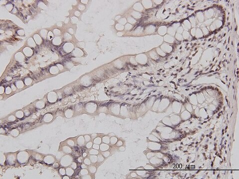
immunohistochemistry: suitable
immunoprecipitation (IP): suitable
western blot: suitable

isotype

IgG1

NCBI accession no.

UniProt accession no.

shipped in

wet ice

target post-translational modification

unmodified

Gene Information

human ... GABRA1(2554)

Specificity

Reacts with the alpha 1-chain of the GABAA receptor in bovine and human but not rat

Immunogen

Epitope: N-terminus
Purified GABA/benzodiazepine receptor from bovine cortex.

Application

Anti-GABA A Receptor α1 chain Antibody, N-terminus, clone BD24 is an antibody against GABA A Receptor α1 chain for use in IH, IP & WB.
Immunohistochemistry: 10-20 μg/mL * See protocol below.

Western blot: 20 μg/mL

Immunoprecipitation

Optimal working dilutions must be determined by end user.

APPLICATION NOTES FOR MAB339

IMMUNOHISTOCHEMISTRY

1) Fresh tissue (human) should be used (3-6 hours postmortem). The tissue should be immersion fixed in 2% paraformaldehyde/0.1% glutaraldehyde. Prepare 50 mm sections and store in cryoprotective solution at -15°C.

2) Sections should be incubated floating in suitable small vials.

3) Block endogenous peroxidase with 100 mmol/L Tris-HCl, 150 mmol/L NaCl, pH 7.4 (Buffer A) containing 3% H2O2 (v/v) and 10% methanol for 20 min at room temperature.

4) Wash sections in Buffer A. Incubate sections in Buffer A containing 5% fetal bovine serum (v/v) and 0.1-0.5% Triton X-100 (v/v).

5) Wash sections in Buffer A. Incubate sections with MAB339 (diluted 10-20 mg/mL in Buffer A containing 5% fetal bovine serum (v/v) and 0.1-0.5% Triton X-100 (v/v) for 12-36 hours at +4°C

6) Wash sections in Buffer A.

7) Detect with standard secondary antibody detection system (PAP, ABC, etc.).

8) Wash sections in Buffer A.

9) Mount sections on chrome alum-coated slides, dry, dehydrate, and apply coverslips.
Research Category
Neuroscience
Research Sub Category
Neurotransmitters & Receptors

Physical form

Format: Purified
Liquid. Buffer = 0.02M Phosphate buffer, 0.25M NaCl with 0.1% sodium azide.

Storage and Stability

Maintain lyophilized material at +2-8°C for up to 12 months. After reconstitution maintain frozen at -20°C in undiluted aliquots for up to 6 months. Avoid repeated freeze/thaw cycles.

Other Notes

Concentration: Please refer to the Certificate of Analysis for the lot-specific concentration.

Legal Information

CHEMICON is a registered trademark of Merck KGaA, Darmstadt, Germany

Disclaimer

Unless otherwise stated in our catalog or other company documentation accompanying the product(s), our products are intended for research use only and are not to be used for any other purpose, which includes but is not limited to, unauthorized commercial uses, in vitro diagnostic uses, ex vivo or in vivo therapeutic uses or any type of consumption or application to humans or animals.

Not finding the right product?  

Try our Product Selector Tool.

Storage Class Code

10 - Combustible liquids

WGK

WGK 2

Flash Point(F)

Not applicable

Flash Point(C)

Not applicable


Certificates of Analysis (COA)

Search for Certificates of Analysis (COA) by entering the products Lot/Batch Number. Lot and Batch Numbers can be found on a product’s label following the words ‘Lot’ or ‘Batch’.

Already Own This Product?

Find documentation for the products that you have recently purchased in the Document Library.

Visit the Document Library

Ivan Milenkovic et al.
Brain structure & function, 223(3), 1501-1518 (2017-11-24)
The function, regulation and cellular distribution of GABAA receptor subunits have been extensively documented in the adult rodent brain and are linked to numerous neurological disorders. However, there is a surprising lack of knowledge on the cellular (sub-) distribution of
Raunak Sinha et al.
Cell, 168(3), 413-426 (2017-01-28)
The fovea is a specialized region of the retina that dominates the visual perception of primates by providing high chromatic and spatial acuity. While the foveal and peripheral retina share a similar core circuit architecture, they exhibit profound functional differences
Lindsey McCracken et al.
Neurology(R) neuroimmunology & neuroinflammation, 4(6), e404-e404 (2017-10-28)
We tested whether antibody screening samples of patients with suspected autoimmune encephalitis with additional research assays would improve the detection of autoimmune encephalitis compared with standard clinical testing alone. We examined 731 samples (333 CSF, 182 sera, and 108 pairs)
Wen-Yi Lo et al.
Neurochemical research, 39(6), 1088-1103 (2013-11-12)
GABAA receptors, the major mediators of fast inhibitory neuronal transmission, are heteropentameric glycoproteins assembled from a panel of subunits, usually including α and β subunits with or without a γ2 subunit. The α1β2γ2 receptor is the most abundant GABAA receptor
Vaishali S Janve et al.
Annals of neurology, 79(5), 806-825 (2016-03-08)
The Epi4K Consortium recently identified 4 de novo mutations in the γ-aminobutyric acid type A (GABAA ) receptor β3 subunit gene GABRB3 and 1 in the β1 subunit gene GABRB1 in children with one of the epileptic encephalopathies (EEs) Lennox-Gastaut

Our team of scientists has experience in all areas of research including Life Science, Material Science, Chemical Synthesis, Chromatography, Analytical and many others.

Contact Technical Service