Accéder au contenu
Merck
Toutes les photos(1)

Documents

MAB2148-C

Sigma-Aldrich

Anti-PECAM-1 Antibody, clone P2B1 (Ascites Free)

clone P2B1, 1 mg/mL, from mouse

Synonyme(s) :

Platelet endothelial cell adhesion molecule, PECAM-1, EndoCAM, GPIIA′, PECA1, CD31

Se connecterpour consulter vos tarifs contractuels et ceux de votre entreprise/organisme


About This Item

Code UNSPSC :
12352203
eCl@ss :
32160702
Nomenclature NACRES :
NA.41

Source biologique

mouse

Niveau de qualité

Forme d'anticorps

purified antibody

Type de produit anticorps

primary antibodies

Clone

P2B1, monoclonal

Espèces réactives

human

Concentration

1 mg/mL

Technique(s)

ELISA: suitable
flow cytometry: suitable
immunocytochemistry: suitable
immunofluorescence: suitable
immunohistochemistry: suitable
immunoprecipitation (IP): suitable

Isotype

IgG1κ

Numéro d'accès NCBI

Numéro d'accès UniProt

Conditions d'expédition

wet ice

Modification post-traductionnelle de la cible

unmodified

Informations sur le gène

human ... PECAM1(5175)

Description générale

PECAM-1 is a type I integral membrane glycoprotein, and a member of the immunoglobulin superfamily of cell surface receptors. PECAM-1 is constitutively expressed on the surface of endothelial cells where it is concentrated at cell junctions. PECAM-1 is also expressed at low levels on all leukocytes and platelets. Homotypic interaction of CD31 is important in cell-cell interaction, cell-matrix interaction and signal transduction.

Immunogène

Whole cells expressing human PECAM-1.

Application

Immunocytochemistry Analysis: A representative lot from an independent laboratory detected PECAM-1 in pluripotent cells (Boyd, N. L., et al. (2007). Exp Biol Med. 232(6):833-843.).

Immunoprecipitation Analysis: A representative lot detected PECAM-1 by immunoprecipitation.

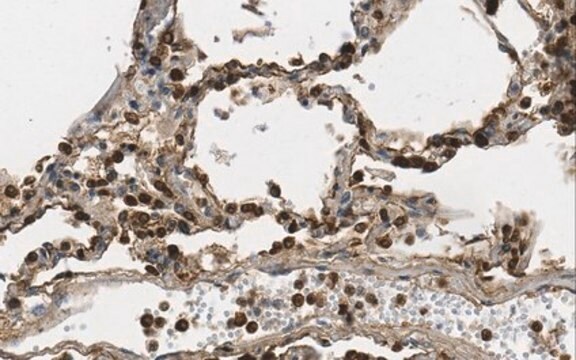
Immunohistochemistry Analysis: A representative lot from an independent laboratory detected PECAM-1 in astrocytoma tissue sections (Bronger, H., et al. (2005). Cancer Res. 65(24):11419-28.).

ELISA Analysis: A representative lot of from an independent laboratory detected PECAM-1 in a panel of CD31 mutant cell lines (Newton, J. P, et al. (1997) J. Biol. Chem., 272: 20555-63.).

Immunofluorescence Analysis: A representative lot from an independent laboratory detected PECAM-1 in astrocytoma tissue sections (Bronger, H., et al. (2005). Cancer Res. 65(24):11419-28.).
Research Category
Cell Structure
Research Sub Category
ECM Proteins
This Anti-PECAM-1 Antibody, clone P2B1 (Ascites Free) is validated for use in flow cytometry, ICC, IP, IHC, ELISA & immunofluorescence for the detection of PECAM-1.

Qualité

Evaluated by Flow Cytometry in human PBMCs.

Flow Cytometry Analysis: 1 µg of this antibody detected PECAM-1 in 1X10E6 human PBMCs.

Description de la cible

~80 kDa calculated

Liaison

Replaces: 04-1074, MAB2148

Forme physique

Format: Purified
Protein G Purified
Purified mouse monoclonal IgG1κ in buffer containing 0.1 M Tris-Glycine (pH 7.4), 150 mM NaCl with 0.05% sodium azide.

Stockage et stabilité

Stable for 1 year at 2-8°C from date of receipt.

Clause de non-responsabilité

Unless otherwise stated in our catalog or other company documentation accompanying the product(s), our products are intended for research use only and are not to be used for any other purpose, which includes but is not limited to, unauthorized commercial uses, in vitro diagnostic uses, ex vivo or in vivo therapeutic uses or any type of consumption or application to humans or animals.

Not finding the right product?  

Try our Outil de sélection de produits.

Code de la classe de stockage

12 - Non Combustible Liquids

Classe de danger pour l'eau (WGK)

WGK 1

Point d'éclair (°F)

Not applicable

Point d'éclair (°C)

Not applicable


Certificats d'analyse (COA)

Recherchez un Certificats d'analyse (COA) en saisissant le numéro de lot du produit. Les numéros de lot figurent sur l'étiquette du produit après les mots "Lot" ou "Batch".

Déjà en possession de ce produit ?

Retrouvez la documentation relative aux produits que vous avez récemment achetés dans la Bibliothèque de documents.

Consulter la Bibliothèque de documents

Laszlo Halasz et al.
International journal of molecular sciences, 25(1) (2024-01-11)
The genome of human adipose-derived stem cells (ADSCs) from abdominal and gluteofemoral adipose tissue depots are maintained in depot-specific stable epigenetic conformations that influence cell-autonomous gene expression patterns and drive unique depot-specific functions. The traditional approach to explore tissue-specific transcriptional
Lei Huang et al.
Nature communications, 8(1), 68-68 (2017-07-14)
Browning of subcutaneous white fat (iWAT) involves several reprograming events, but the underlying mechanisms are incompletely understood. Here we show that the transcription factor Hlx is selectively expressed in brown adipose tissue (BAT) and iWAT, and is translationally upregulated by

Notre équipe de scientifiques dispose d'une expérience dans tous les secteurs de la recherche, notamment en sciences de la vie, science des matériaux, synthèse chimique, chromatographie, analyse et dans de nombreux autres domaines..

Contacter notre Service technique