Skip to Content
Merck
All Photos(3)

Documents

ABN78

Sigma-Aldrich

Anti-NeuN Antibody (rabbit)

from rabbit, purified by affinity chromatography

Synonym(s):

RNA binding protein fox-1 homolog 3, Fox-1 homolog C, Fox-3, Hexaribonucleotide-binding protein 3, NeuN, Neuronal nuclei

Sign Into View Organizational & Contract Pricing


About This Item

UNSPSC Code:
12352203
eCl@ss:
32160702
NACRES:
NA.41

biological source

rabbit

Quality Level

antibody form

affinity isolated antibody

antibody product type

primary antibodies

clone

polyclonal

purified by

affinity chromatography

species reactivity

mouse, human, snail, rat

technique(s)

immunocytochemistry: suitable
immunofluorescence: suitable
immunohistochemistry: suitable
western blot: suitable

NCBI accession no.

UniProt accession no.

shipped in

wet ice

storage temp.

2-8°C

target post-translational modification

unmodified

Gene Information

General description

RNA binding protein fox-1 homolog 3 also known as NeuN (Neuronal nuclei) is encoded by the Rbfox3 (also known as D11Bwg0517e, Hrnbp3) gene (Gene ID 52897) in murine species. The RNA-binding Fox (Rbfox) family of splicing factors is comprised of three members, Rbfox1 (Fox-1 or A2BP1), Rbfox2 (Fox-2 or RBM9), and Rbfox3 (Fox-3, HRNBP3 or NeuN). Rbfox proteins regulate splicing of many neuronal transcripts (pre-mRNAs) by binding the sequence (U)GCAUG in introns flanking alternative exons. A (U)GCAUG motif that lies downstream of the alternative exon generally promotes Rbfox-dependent exon inclusion, whereas an upstream motif will usually repress exon inclusion. Originally characterized as a marker of post-mitotic neurons and named neuronal nuclei (NeuN), Rbfox3 plays a role in promoting neuronal differentiation through alternative splicing of Numb pre-mRNA during brain development. Rbfox3-knockout in mice is reported to result in defective hippocampal gene expression, defects in synaptic transmission and plasticity in the dentate gyrus, as well as increased seizure susceptibility and decreased anxiety-related behaviors. Likewise, human RBFOX3 gene mutations have been linked to neurodevelopmental delay, cognitive impairments, autistic features, and epilepsy.

Specificity

Anti NeuN antibody from rabbit is a polyclonal antibody that targets NeuN N-terminal region that ends at the start of the RNA recognition motif (RRM). The target region is present in all human, mouse, and rat spliced isoforms reported by UniProt (A6NFN3, Q8BIF2, D4A2H6, A0A0G2K2J4).

Immunogen

Epitope: N-terminus
GST-tagged recombinant mouse NeuN N-terrminal fragment.

Application

Anti-NeuN (rabbit) Antibody, Cat. No. ABN78, is a highly specific rabbit polyclonal antibody that targets NeuN and has been tested in Immunocytochemistry, Immunofluorescence, Immunohistochemistry, and Western Blotting.
Immunocytochemistry Analysis: A 1:500 dilution from a representative lot detected NeuN in cultured embryonic E18 rat hippocampus neurons.

Immunohistochemistry Analysis: A 1:500 dilution from a representative lot detected NeuN-positive neurons in human (cerebellum and cerebral cortex) and mouse (hippocampus) brain tissue sections.

Immunocytochemistry Analysis: A representative lots immunostained 4% paraformaldehyde-fixed neurons isolated from snail brain (Nikolić, L., et al. (2013). J. Exp. Biol. 216(Pt 18):3531-3541).

Immunofluorescence Analysis: Representative lots detected NeuN-positive neurons in frozen brain sections from 4% paraformaldehyde-perfused mice (Cherry, J.D., et al. (2015). J. Neuroinflammation. 12:203; Huang, B., et al. (2015). Neuron. 85(6):1212-1226; Ataka, K., et al. (2013). PLoS One. 8(11):e81744).

Immunofluorescence Analysis: A representative lots detected NeuN-positive neurons in formalin-fixed and paraffin-embedded amyotrophic lateral sclerosis (ALS) human brain tissue sections (Fratta, P., et al. (2015). Neurobiol. Aging. 36(1):546.e1-7).

Immunohistochemistry Analysis: A representative lot detected NeuN-positive neurons in rat brain tissue sections (Mendonça, M.C., et al. (2013).Toxins. 5(12):2572-2588).

Western Blotting Analysis: Representative lot detected NeuN in mouse and rat brain tissue lysates (Huang, B., et al. (2015). Neuron. 85(6):1212-1226; Mendonça, M.C., et al. (2013).Toxins. 5(12):2572-2588).

This rabbit polyclonal antibody is also avaiable as Alexa Fluor 488 (ABN78A4), biotin (ABN78B), and Cy3 (ABN78C3) conjugates for immunocytochemistry, immunofluorescence, immunohistochemistry applications.
Research Category
Neuroscience
Research Sub Category
Developmental Signaling

Quality

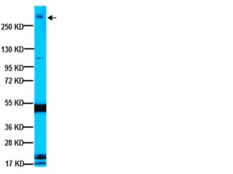
Evaluated by Western Blotting in mouse brain nuclear extract.

Western Blotting Analysis: 0.2 µg/mL of this antibody detected NeuN in 10 µg of mouse brain nuclear extract.

Target description

~48/42 kDa observed. 33.87/35.12 kDa (human isoform 1/2), 40.61/39.05/35.64/34.07/35.63 kDa (mouse isoform 1/2/3/4/5), 40.59kDa (rat; UniProt D4A2H6), 34.05 kDa (rat; UniProt A0A0G2K2J4) calculated. Uncharacterized bands may be observed in some lysate(s).

Physical form

Affinity purified
Purified rabbit polyclonal antibody in buffer containing 0.1 M Tris-Glycine (pH 7.4), 150 mM NaCl with 0.05% sodium azide.

Storage and Stability

Stable for 1 year at 2-8°C from date of receipt.

Analysis Note

Control
Mouse brain nuclear extract

Other Notes

Concentration: Please refer to lot specific datasheet.

Legal Information

ALEXA FLUOR is a trademark of Life Technologies

Disclaimer

Unless otherwise stated in our catalog or other company documentation accompanying the product(s), our products are intended for research use only and are not to be used for any other purpose, which includes but is not limited to, unauthorized commercial uses, in vitro diagnostic uses, ex vivo or in vivo therapeutic uses or any type of consumption or application to humans or animals.

Not finding the right product?  

Try our Product Selector Tool.

Storage Class Code

12 - Non Combustible Liquids

WGK

WGK 1

Flash Point(F)

Not applicable

Flash Point(C)

Not applicable


Certificates of Analysis (COA)

Search for Certificates of Analysis (COA) by entering the products Lot/Batch Number. Lot and Batch Numbers can be found on a product’s label following the words ‘Lot’ or ‘Batch’.

Already Own This Product?

Find documentation for the products that you have recently purchased in the Document Library.

Visit the Document Library

C9orf72 frontotemporal lobar degeneration is characterised by frequent neuronal sense and antisense RNA foci.
Mizielinska, S; Lashley, T; Norona, FE; Clayton, EL; Ridler, CE; Fratta, P; Isaacs, AM
Acta neuropathologica null
Jeong-Hwi Cho et al.
Molecular medicine reports, 13(4), 2949-2956 (2016-03-05)
4-Hydroxy‑3-methoxybenzaldehyde (vanillin) and 4-hydroxybenzyl alcohol (4-HBA) are well‑known phenolic compounds, which possess various therapeutic properties and are widely found in a variety of plants. In the present study, the effects of vanillin and 4‑HBA were first investigated on cell proliferation
Félix Leroy et al.
eLife, 3, doi:10-doi:10 (2014-10-15)
In amyotrophic lateral sclerosis (ALS) the large motoneurons that innervate the fast-contracting muscle fibers (F-type motoneurons) are vulnerable and degenerate in adulthood. In contrast, the small motoneurons that innervate the slow-contracting fibers (S-type motoneurons) are resistant and do not degenerate.
Monique C P Mendonça et al.
Toxins, 5(12), 2572-2588 (2013-12-20)
Apart from its angiogenic and vascular permeation activity, the vascular endothelial growth factor (VEGF) has been also reported as a potent neuronal protector. Newborn rats with low VEGF levels develop neuron degeneration, while high levels induce protective mechanisms in several
Rachel E Bennett et al.
Journal of neurotrauma, 31(19), 1647-1663 (2014-05-07)
The pathological processes that lead to long-term consequences of multiple concussions are unclear. Primary mechanical damage to axons during concussion is likely to contribute to dysfunction. Secondary damage has been hypothesized to be induced or exacerbated by inflammation. The main

Articles

Explore the basics of working with antibodies including technical information on structure, classes, and normal immunoglobulin ranges.

Learn differences in monoclonal vs polyclonal antibodies including how antibodies are generated, clone numbers, and antibody formats.

Immunofluorescence uses antibody-conjugated fluorescent molecules for protein localization, modification confirmation, and protein complex visualization.

Protocols

Tips and troubleshooting for FFPE and frozen tissue immunohistochemistry (IHC) protocols using both brightfield analysis of chromogenic detection and fluorescent microscopy.

Our team of scientists has experience in all areas of research including Life Science, Material Science, Chemical Synthesis, Chromatography, Analytical and many others.

Contact Technical Service