
AB2047
Anti-Fibronectin Antibody
Chemicon®, from rabbit
About This Item
Productos recomendados


origen biológico
rabbit
forma del anticuerpo
affinity purified immunoglobulin
tipo de anticuerpo
primary antibodies
clon
polyclonal
reactividad de especies
bovine
fabricante / nombre comercial
Chemicon®
técnicas
ELISA: suitable
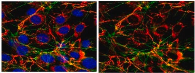
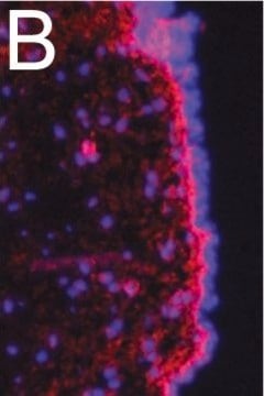
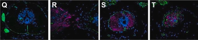
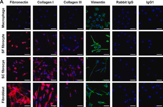
immunofluorescence: suitable
immunohistochemistry: suitable (paraffin)
radioimmunoassay: suitable
western blot: suitable
Nº de acceso UniProt
Condiciones de envío
dry ice
modificación del objetivo postraduccional
unmodified
Descripción general
Fibronectin exists in two main forms: 1) as an insoluble glycoprotein dimer that serves as a linker in the ECM (extracellular matrix), and; 2) as a soluble disulphide linked dimer found in the plasma (plasma FN). The plasma form is synthesized by hepatocytes, and the ECM form is made by fibroblasts, chondrocytes, endothelial cells, macrophages, as well as certain epithelial cells.
Fibronectin sometimes serves as a general cell adhesion molecule by anchoring cells to collagen or proteoglycan substrates. FN also can serve to organize cellular interaction with the ECM by binding to different components of the extracellular matrix and to membrane-bound FN receptors on cell surfaces. The importance of fibronectin in cell migration events during embryogenesis has been documented in several contexts, e.g.: 1) mesodermal cell migration during gastrulation can be blocked by injection of Arg-Gly-Asp (RGD) tripeptides that block cellular FN receptors (integrins); 2) injection of anti-FN antibodies into chick embryos blocks migration of precardiac cells to the embryonic midline, and; 3) the patterns of FN deposition in developing vertebrate limbs determines the patterns of precartilage cell adhesion to the ECM, thereby specifying limb-specific patterns of chondrogenesis. {D. Marcey, http://www.clunet.edu/BioDev/omm/fibro/frames/fibrotxt.htm}.
Especificidad
Inmunógeno
Aplicación
Immunohistochemistry on paraffin embedded tissues requires light fixation in 2% PFA, 4% formalin (less than 90 minutes), acetone or methyl-carnoy fixation; traditional formalin fixation is not recommended. Antigen retrieval is HIER citrate buffer; detection is via enhanced enzymatic methods only.
Immunoblotting: 1:1000 of 2% deoxycholate + 10% SDS, 6M urea extracted bovine cell cultures (Kinsella, 2000). Antibody demonstates the appropriate twin bands at ~220kDa.
Radioimmunoassay
ELISA
Optimal working dilutions must be determined by end user.
Cell Structure
ECM Proteins
Forma física
Almacenamiento y estabilidad
Otras notas
Información legal
Cláusula de descargo de responsabilidad
¿No encuentra el producto adecuado?
Pruebe nuestro Herramienta de selección de productos.
Código de clase de almacenamiento
12 - Non Combustible Liquids
Clase de riesgo para el agua (WGK)
WGK 2
Punto de inflamabilidad (°F)
Not applicable
Punto de inflamabilidad (°C)
Not applicable
Certificados de análisis (COA)
Busque Certificados de análisis (COA) introduciendo el número de lote del producto. Los números de lote se encuentran en la etiqueta del producto después de las palabras «Lot» o «Batch»
¿Ya tiene este producto?
Encuentre la documentación para los productos que ha comprado recientemente en la Biblioteca de documentos.
Filtros activos
Nuestro equipo de científicos tiene experiencia en todas las áreas de investigación: Ciencias de la vida, Ciencia de los materiales, Síntesis química, Cromatografía, Analítica y muchas otras.
Póngase en contacto con el Servicio técnico