Accéder au contenu
Merck
Toutes les photos(2)

Key Documents

MABN11

Sigma-Aldrich

Anti-Amyloid β40 Antibody, clone G2-10

clone G2-10, from mouse

Synonyme(s) :

Alzheimer disease, Alzheimer disease amyloid protein, Cerebral vascular amyloid peptide, Protease nexin-II, amyloid beta (A4) precursor protein, amyloid beta A4 protein, amyloid beta precursor protein, beta-amyloid peptide, human mRNA for amyloid A4 prec

Se connecterpour consulter vos tarifs contractuels et ceux de votre entreprise/organisme


About This Item

Code UNSPSC :
12352203
eCl@ss :
32160702
Nomenclature NACRES :
NA.41

Source biologique

mouse

Niveau de qualité

Forme d'anticorps

purified antibody

Clone

G2-10, monoclonal

Espèces réactives

human, mouse

Technique(s)

ELISA: suitable
immunohistochemistry: suitable
western blot: suitable

Isotype

IgG2bκ

Numéro d'accès NCBI

Numéro d'accès UniProt

Conditions d'expédition

wet ice

Modification post-traductionnelle de la cible

unmodified

Informations sur le gène

human ... APP(351)

Description générale

The cerebral and vascular plaques associated with Alzheimer′s disease (AD) are mainly composed of amyloid beta peptides (Aβ). Aβ is derived from cleavage of the amyloid precursor protein (APP) and varies in length from 39 to 43 amino acids. Aβ [1-40], Aβ [1-42], and Aβ [1-43] peptides result from cleavage of APP after residues 40, 42, and 43, respectively. The cleavage takes place by gamma-secretase during the last APP processing step. Aβ [1-40], [1-42] and [1-43] peptides are major constituents of the plaques and tangles that occur in AD. Aβ antibodies and peptides have been developed as tools for elucidating the biology of AD.

Spécificité

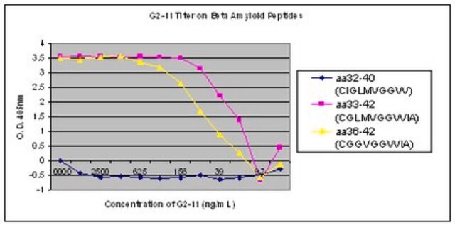
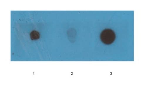
This antibody recognizes Amyloid β40 at the C-terminus.

Immunogène

Epitope: C-terminus
Linear peptide of human Amyloid β40 at the C terminus.

Application

Detect Amyloid β40 using this Anti-Amyloid β40 Antibody, clone G2-10 validated for use in WB, IH, ELISA.
Immunohistochemistry Analysis: 1:300 dilution from a previous lot detected Amyloid β40 in Alzheimer’s diseased hippocampus tissue.
Research Category
Neuroscience
Research Sub Category
Neurodegenerative Diseases

Qualité

Evaluated by Western Blot in APP transgenic CRND8 mouse brain lysate.

Western Blot Analysis: 1 µg/ml of this antibody detected Amyloid β40 on 10 µg of APP transgenic CRND8 mouse brain lysate.

Description de la cible

~ 4 kDa

Forme physique

Format: Purified
Protein G Purified
Purified mouse monoclonal IgG2bκ in buffer containing 0.1 M Tris-Glycine (pH 7.4, 150 mM NaCl) with 0.05% sodium azide and 1% BSA.

Stockage et stabilité

Stable for 1 year at 2-8°C from date of receipt.

Remarque sur l'analyse

Control
APP transgenic CRND8 mouse brain lysate

Autres remarques

Concentration: Please refer to the Certificate of Analysis for the lot-specific concentration.

Clause de non-responsabilité

Unless otherwise stated in our catalog or other company documentation accompanying the product(s), our products are intended for research use only and are not to be used for any other purpose, which includes but is not limited to, unauthorized commercial uses, in vitro diagnostic uses, ex vivo or in vivo therapeutic uses or any type of consumption or application to humans or animals.

Code de la classe de stockage

12 - Non Combustible Liquids

Classe de danger pour l'eau (WGK)

WGK 1

Point d'éclair (°F)

Not applicable

Point d'éclair (°C)

Not applicable


Certificats d'analyse (COA)

Recherchez un Certificats d'analyse (COA) en saisissant le numéro de lot du produit. Les numéros de lot figurent sur l'étiquette du produit après les mots "Lot" ou "Batch".

Déjà en possession de ce produit ?

Retrouvez la documentation relative aux produits que vous avez récemment achetés dans la Bibliothèque de documents.

Consulter la Bibliothèque de documents

Ditte Z Christensen et al.
Frontiers in aging neuroscience, 6, 139-139 (2014-07-16)
Abnormalities and impairments in axonal transport are suggested to strongly contribute to the pathological alterations underlying AD. The exact mechanisms leading to axonopathy are currently unclear, but it was recently suggested that APP expression itself triggers axonal degeneration. We used
Ge Li et al.
International journal of molecular medicine, 42(4), 1935-1944 (2018-08-08)
Aging is associated with impairment of the paravascular pathway caused by the activation of astrocytes and depolarization of protein aquaporin‑4 (AQP4) water channels, resulting in the accumulation of protein waste, including amyloid β (Aβ), in the brain parenchyma. The secreted
Kevin Kleffman et al.
Cancer discovery, 12(5), 1314-1335 (2022-03-10)
Brain metastasis is a significant cause of morbidity and mortality in multiple cancer types and represents an unmet clinical need. The mechanisms that mediate metastatic cancer growth in the brain parenchyma are largely unknown. Melanoma, which has the highest rate
Jin Cui et al.
Cell discovery, 1, 15021-15021 (2015-01-01)
Despite decades of intense global effort, no disease-modifying drugs for Alzheimer's disease have emerged. Molecules targeting catalytic activities of γ-secretase or β-site APP-cleaving enzyme 1 (BACE1) have been beset by undesired side effects. We hypothesized that blocking the interaction between
Vanessa Kurth et al.
The Journal of biological chemistry, 299(8), 104997-104997 (2023-07-03)
Presenilin-1 (PSEN1) is the catalytic subunit of the intramembrane protease γ-secretase and undergoes endoproteolysis during its maturation. Heterozygous mutations in the PSEN1 gene cause early-onset familial Alzheimer's disease (eFAD) and increase the proportion of longer aggregation-prone amyloid-β peptides (Aβ42 and/or

Notre équipe de scientifiques dispose d'une expérience dans tous les secteurs de la recherche, notamment en sciences de la vie, science des matériaux, synthèse chimique, chromatographie, analyse et dans de nombreux autres domaines..

Contacter notre Service technique