Accéder au contenu
Merck
Toutes les photos(3)

Principaux documents

MABE288

Sigma-Aldrich

Anti-PCNA Antibody, clone PC10

clone PC10, from mouse

Synonyme(s) :

Proliferating cell nuclear antigen, PCNA, Cyclin

Se connecterpour consulter vos tarifs contractuels et ceux de votre entreprise/organisme


About This Item

Code UNSPSC :
12352203
eCl@ss :
32160702
Nomenclature NACRES :
NA.41

Source biologique

mouse

Niveau de qualité

Forme d'anticorps

purified immunoglobulin

Type de produit anticorps

primary antibodies

Clone

PC10, monoclonal

Espèces réactives

human

Technique(s)

immunofluorescence: suitable
immunohistochemistry: suitable
western blot: suitable

Isotype

IgG2aκ

Numéro d'accès NCBI

Numéro d'accès UniProt

Conditions d'expédition

wet ice

Modification post-traductionnelle de la cible

unmodified

Informations sur le gène

human ... PCNA(5111)

Description générale

Proliferating cell nuclear antigen (PCNA) is a heterotrimeric nuclear protein that functions as a clamp on replicating DNA. In cases of DNA damage and failure of the DNA repair pathways, PCNA mediates translesion DNA synthesis, a DNA damage tolerance (DDT) pathway, which allows replication to progress in spite of large lesions to DNA which may be caused by replication stress, ionizing radiation, spontaneous DNA damage, among other factors. PCNA primarily recruits low-fidelity DNA polymerase enzymes that are capable of elongating DNA over lesion sites. PCNA is regulated by cycles of ubiquitination, depending on the DDT pathway involved: PCNA is monoubiquitinated by Rad6–Rad18; polyubiquitinated by the Mms2-Ubc13-Rad5; or sumoylated by the Ubc9-Siz1 complex.

Immunogène

Recombinant protein corresponding to human PCNA.

Application

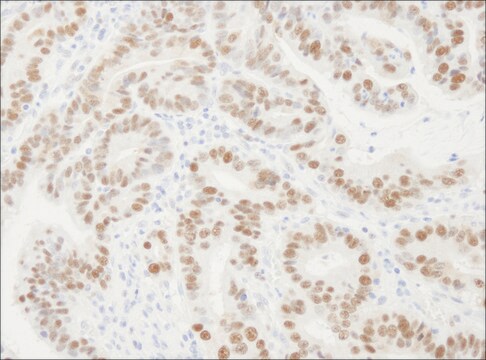
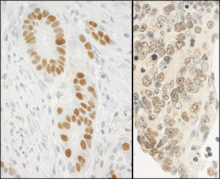
Immunohistochemistry Analysis: A 1:1,000 dilution from a representative lot detected PCNA in human colon adenocarcinoma tissues and human tonsil tissues.

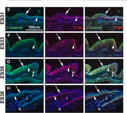
Immunofluorescence Analysis: A 1:1,500 dilution from a representative lot detected PCNA in human lymph nodes and in human colon adenocarcinoma cells.
Research Category
Epigenetics & Nuclear Function
Research Sub Category
Cell Cycle, DNA Replication & Repair
Use Anti-PCNA Antibody, clone PC10 (Mouse Monoclonal Antibody) validated in WB, IHC, IF to detect PCNA also known as Proliferating cell nuclear antigen, PCNA, Cyclin.

Qualité

Evaluated by Western Blot in HeLa cell lysate.

Western Blot Analysis: 0.01 µg/mL of this antibody recognized PCNA in 10 µg of HeLa cell lysate.

Description de la cible

~29 kDa observed

Forme physique

Format: Purified
Protein G Purified
Purified mouse monoclonal IgG2aκ in buffer containing 0.1 M Tris-Glycine (pH 7.4), 150 mM NaCl with 0.05% sodium azide.

Stockage et stabilité

Stable for 1 year at 2-8°C from date of receipt.

Remarque sur l'analyse

Control
HeLa cell lysate

Autres remarques

Concentration: Please refer to the Certificate of Analysis for the lot-specific concentration.

Clause de non-responsabilité

Unless otherwise stated in our catalog or other company documentation accompanying the product(s), our products are intended for research use only and are not to be used for any other purpose, which includes but is not limited to, unauthorized commercial uses, in vitro diagnostic uses, ex vivo or in vivo therapeutic uses or any type of consumption or application to humans or animals.

Vous ne trouvez pas le bon produit ?  

Essayez notre Outil de sélection de produits.

Code de la classe de stockage

12 - Non Combustible Liquids

Classe de danger pour l'eau (WGK)

WGK 1

Point d'éclair (°F)

Not applicable

Point d'éclair (°C)

Not applicable


Certificats d'analyse (COA)

Recherchez un Certificats d'analyse (COA) en saisissant le numéro de lot du produit. Les numéros de lot figurent sur l'étiquette du produit après les mots "Lot" ou "Batch".

Déjà en possession de ce produit ?

Retrouvez la documentation relative aux produits que vous avez récemment achetés dans la Bibliothèque de documents.

Consulter la Bibliothèque de documents

Sarah Watson et al.
Cell reports, 42(1), 112013-112013 (2023-01-20)
Clinical sequencing efforts are rapidly identifying sarcoma gene fusions that lack functional validation. An example is the fusion of transcriptional coactivators, VGLL2-NCOA2, found in infantile rhabdomyosarcoma. To delineate VGLL2-NCOA2 tumorigenic mechanisms and identify therapeutic vulnerabilities, we implement a cross-species comparative
Rindert Missiaen et al.
Cell metabolism, 34(8), 1151-1167 (2022-07-16)
Hepatocellular carcinoma (HCC) is a typically fatal malignancy exhibiting genetic heterogeneity and limited therapy responses. We demonstrate here that HCCs consistently repress urea cycle gene expression and thereby become auxotrophic for exogenous arginine. Surprisingly, arginine import is uniquely dependent on
Hai-Yan Wu et al.
NPJ Regenerative medicine, 6(1), 36-36 (2021-07-01)
Cardiovascular disease is the leading cause of death in the world due to losing regenerative capacity in the adult heart. Frogs possess remarkable capacities to regenerate multiple organs, including spinal cord, tail, and limb, but the response to heart injury
Helen Eachus et al.
Communications biology, 7(1), 416-416 (2024-04-06)
Exposure to excess glucocorticoid (GC) during early development is implicated in adult dysfunctions. Reduced adult hippocampal neurogenesis is a well-known consequence of exposure to early life stress or elevated GC, however the effects on neurogenesis during development and effects on
Michalis Petropoulos et al.
Nature, 628(8007), 433-441 (2024-03-21)
An important advance in cancer therapy has been the development of poly(ADP-ribose) polymerase (PARP) inhibitors for the treatment of homologous recombination (HR)-deficient cancers1-6. PARP inhibitors trap PARPs on DNA. The trapped PARPs are thought to block replisome progression, leading to

Articles

Loading controls in western blotting application.

Notre équipe de scientifiques dispose d'une expérience dans tous les secteurs de la recherche, notamment en sciences de la vie, science des matériaux, synthèse chimique, chromatographie, analyse et dans de nombreux autres domaines..

Contacter notre Service technique