Accéder au contenu
Merck
Toutes les photos(4)

Key Documents

ABN14

Sigma-Aldrich

Anti-Brain lipid binding protein Antibody

from rabbit, purified by affinity chromatography

Synonyme(s) :

Brain lipid-binding protein, Fatty acid-binding protein 7, Mammary-derived growth inhibitor related, brain lipid binding protein, fatty acid binding protein 7, brain, mammary-derived growth inhibitor-related

Se connecterpour consulter vos tarifs contractuels et ceux de votre entreprise/organisme


About This Item

Code UNSPSC :
12352203
eCl@ss :
32160702
Nomenclature NACRES :
NA.41

Source biologique

rabbit

Niveau de qualité

Forme d'anticorps

affinity isolated antibody

Type de produit anticorps

primary antibodies

Clone

polyclonal

Produit purifié par

affinity chromatography

Espèces réactives

mouse

Réactivité de l'espèce (prédite par homologie)

human (based on 100% sequence homology)

Conditionnement

antibody small pack of 25 μg

Technique(s)

immunohistochemistry: suitable
western blot: suitable

Numéro d'accès NCBI

Numéro d'accès UniProt

Conditions d'expédition

ambient

Modification post-traductionnelle de la cible

unmodified

Informations sur le gène

human ... FABP7(2173)
mouse ... Fabp7(12140)

Description générale

BLBP (Brain lipid-binding protein) belongs to a family of small, highly conserved, cytoplasmic proteins that bind long-chain fatty acids and other hydrophobic ligands. BLBPs play essential roles in fatty acid uptake, transport and metabolism. BLBP can be used as a molecular marker for radial glia (a major neural progenitor cell type and a scaffolding supporting neuronal migration) in the developing central nervous system. Radial glia play an essential role in the generation of the cerebral cortex through their function as neuronal precursors and as neuronal migration guides.

Spécificité

Rat homology - 88%.
This antibody recognizes Brain lipid-binding protein.

Immunogène

Epitope: Cytoplasmic Domain
GST-tagged recombinant protein corresponding to human Brain lipid-binding protein.

Application

Detect Brain lipid binding protein using this Anti-Brain lipid binding protein Antibody validated for use in WB, IH.
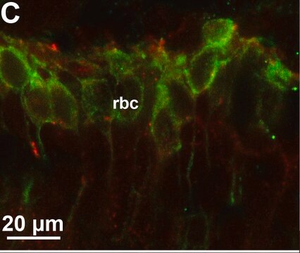
Immunohistochemistry Analysis: A 1:300 dilution of ABN14 antibody from a previous lot detected Brain lipid binding protein in mouse cerebellum tissue.
Research Category
Neuroscience
Research Sub Category
Neuronal & Glial Markers

Qualité

Evaluated by Western Blot in P1 mouse brain tissue lysate.

Western Blot Analysis: 0.01 µg/ml of this antibody detected Brain lipid-binding protein in 10 µg of P1 mouse brain tissue lysate.

Description de la cible

~ 15 kDa

Liaison

Replaces: AB9558

Forme physique

Affinity purified
Purified rabbit polyclonal in buffer containing 0.1 M Tris-Glycine (pH 7.4, 150 mM NaCl) with 0.05% sodium azide.

Stockage et stabilité

Stable for 1 year at 2-8°C from date of receipt.

Remarque sur l'analyse

Control
P1 mouse brain tissue lysate

Autres remarques

Concentration: Please refer to the Certificate of Analysis for the lot-specific concentration.

Clause de non-responsabilité

Unless otherwise stated in our catalog or other company documentation accompanying the product(s), our products are intended for research use only and are not to be used for any other purpose, which includes but is not limited to, unauthorized commercial uses, in vitro diagnostic uses, ex vivo or in vivo therapeutic uses or any type of consumption or application to humans or animals.

Vous ne trouvez pas le bon produit ?  

Essayez notre Outil de sélection de produits.

Code de la classe de stockage

12 - Non Combustible Liquids

Classe de danger pour l'eau (WGK)

WGK 1

Point d'éclair (°F)

Not applicable

Point d'éclair (°C)

Not applicable


Certificats d'analyse (COA)

Recherchez un Certificats d'analyse (COA) en saisissant le numéro de lot du produit. Les numéros de lot figurent sur l'étiquette du produit après les mots "Lot" ou "Batch".

Déjà en possession de ce produit ?

Retrouvez la documentation relative aux produits que vous avez récemment achetés dans la Bibliothèque de documents.

Consulter la Bibliothèque de documents

Constance A Rich et al.
Glia, 66(12), 2617-2631 (2018-09-27)
We and others previously showed that in mouse embryos lacking the transcription factor Sox10, olfactory ensheathing cell (OEC) differentiation is disrupted, resulting in defective olfactory axon targeting and fewer gonadotropin-releasing hormone (GnRH) neurons entering the embryonic forebrain. The underlying mechanisms
A mouse model of human primitive neuroectodermal tumors resulting from microenvironmentally-driven malignant transformation of orthotopically transplanted radial glial cells.
Malchenko, S; Sredni, ST; Hashimoto, H; Kasai, A; Nagayasu, K; Xie, J; Margaryan et al.
Testing null
Vivek K Bajpai et al.
Stem cells (Dayton, Ohio), 35(5), 1402-1415 (2017-02-01)
During development, neural crest (NC) cells are induced by signaling events at the neural plate border of all vertebrate embryos. Initially arising within the central nervous system, NC cells subsequently undergo an epithelial to mesenchymal transition to migrate into the
Sara Anna Bonini et al.
Cerebral cortex (New York, N.Y. : 1991), 26(6), 2832-2849 (2016-03-08)
Alterations in genes that regulate neurodevelopment can lead to cortical malformations, resulting in malfunction during postnatal life. The NF-κB pathway has a key role during neurodevelopment by regulating the maintenance of the neural progenitor cell pool and inhibiting neuronal differentiation.
Mayumi Okamoto et al.
Nature communications, 7, 11349-11349 (2016-04-21)
During cerebral development, many types of neurons are sequentially generated by self-renewing progenitor cells called apical progenitors (APs). Temporal changes in AP identity are thought to be responsible for neuronal diversity; however, the mechanisms underlying such changes remain largely unknown.

Notre équipe de scientifiques dispose d'une expérience dans tous les secteurs de la recherche, notamment en sciences de la vie, science des matériaux, synthèse chimique, chromatographie, analyse et dans de nombreux autres domaines..

Contacter notre Service technique