Direkt zum Inhalt
Merck

MABF972

Sigma-Aldrich

Anti-Complement C3b/iC3b Antibody, clone 3E7, neutralizing

clone 3E7, from mouse

Synonym(e):

Complement C3, C3 and PZP-like alpha-2-macroglobulin domain-containing protein 1, Complement C3 beta chain, C3-beta-c, C3bc, Complement C3 alpha chain, C3a anaphylatoxin, Acylation stimulating protein, ASP, C3adesArg, Complement C3b alpha′ chain, Complem

Anmeldenzur Ansicht organisationsspezifischer und vertraglich vereinbarter Preise


About This Item

UNSPSC-Code:
12352203
eCl@ss:
32160702
NACRES:
NA.43

Biologische Quelle

mouse

Qualitätsniveau

Antikörperform

purified immunoglobulin

Antikörper-Produkttyp

primary antibodies

Klon

3E7, monoclonal

Speziesreaktivität

human, primate

Methode(n)

flow cytometry: suitable
immunocytochemistry: suitable
neutralization: suitable

Isotyp

IgG1κ

NCBI-Hinterlegungsnummer

UniProt-Hinterlegungsnummer

Versandbedingung

wet ice

Posttranslationale Modifikation Target

unmodified

Angaben zum Gen

human ... C3(718)

Allgemeine Beschreibung

Complement C3 (UniProt P01024; also known as C3 and PZP-like alpha-2-macroglobulin domain-containing protein 1) is encoded by the C3 (also known as CPAMD1) gene (Gene ID 718) in human. C3 is initially translated with an N-terminal 22-amino acid signal peptide sequence, which is then removed to produce the 1641-amino acid mature C3. It plays a central role in the activation of the complement system. Its activation is required for the activation of both classical and alternative pathway of complement (CPC and APC, respectively). C3 is cleaved into C3a and C3b during CPC activation by the C3-convertase C4b2a composed of the activated C4 and C2. In APC, C3 is cleaved by the C3-convertase C3bBb composed C3b and the activated form of factor B (Bb). C3b serves as an opsonizing agent, and can be further cleaved by Factor I into C3c and C3d. iC3b is a proteolytically inactive C3b fragment that still opsonizes target microbes or cells, but cannot further amplify/activate the complement cascade through APC. iC3b can be further cleaved to C3dg, and finally to C3d. Unregulated activation of APC can result in paroxysmal nocturnal hemoglobinuria (PNH) that is characterized by chronic intravascular hemolysis. Clinical C5-neutralizing mAb treatment prevents the formation of cytolytic membrane attack complex (MAC) of complement, but does not block APC activation. Consequently, PNH patients are left with immune-mediated hemolytic anemia and their erythrocytes become opsonized with complement C3. Monoclonal antibodies (mAbs) against C3b/iC3b are useful for monitoring and studying C3b/iC3b deposit on PNH blood cells and mAbs with neutralizing activities are useful tools for studying C3-mediated CPC and APC.

Spezifität

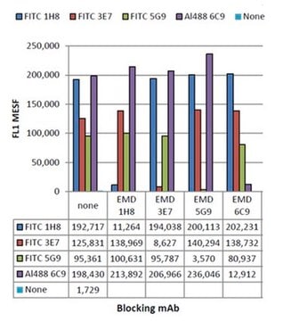
This clone blocks the activation of alternative pathway of complement (APC) by binding C3b and iC3b.

Immunogen

Epitope: iC3b
Sepharose 4B beads with surface C3b/C3bi deposits via APC in normal human serum corresponding to the iC3b of human Complement C3b/iC3b.

Anwendung

Research Category
Entzündung & Immunologie
Research Sub Category
Immunoglobuline & Immunologie
Neutralizing Analysis: This clone has been shown to prevent alternative pathway of complement (APC) activation-mediated hemolysis of IgM-opsinized paroxysmal nocturnal hemoglobinuria (PNH) erthyrocytes (Lindorfer, M.A., et al. (2010). Blood. 115(11):2283-2291).
Flow Cytometry Analysis: A presentive lot detected C3b/iC3b deposit on anti-CD20 mAb Rituximab-opsinized human peripheral blood ARH-77 & Raji B cells, as well as Rituximab-opsinized CD20+ cells freshly isolated from monkey blood (Kennedy, A.D., et al. (2003). Blood. 101(3):1071-1079).
Immunocytochemistry Analysis: A presentive lot detected C3b/iC3b deposit on anti-CD20 mAb Rituximab-opsinized human peripheral blood ARH-77 & Raji B cells, as well as Rituximab-opsinized CD20+ cells freshly isolated from monkey blood (Kennedy, A.D., et al. (2003). Blood. 101(3):1071-1079).
This mouse monoclonal Anti-Complement C3b/iC3b Antibody, clone 3E7, Cat. No. MABF972 is a neutralizing antibody validated for use in Flow Cytometry, for the detection of C3b.

Qualität

Flow Cytometry Analysis: This antibody (200 ug mAb/5 x 10E6 cells/mL) detected C3b/iC3b deposit on human Burkett′s lymphoma Raji B cells opsonized with anti-CD20 mAb Rituximab (RTX) in the presence of 50% normal human serum (NHS).

Zielbeschreibung

187 kDa calculated

Physikalische Form

Protein G Purified
Format: Purified
Purified mouse monoclonal IgG1κ antibody in buffer containing PBS without preservatives.

Lagerung und Haltbarkeit

Stable for 1 year at -20°C from date of receipt.
Handling Recommendations: Upon receipt and prior to removing the cap, centrifuge the vial and gently mix the solution. Aliquot into microcentrifuge tubes and store at -20°C. Avoid repeated freeze/thaw cycles, which may damage IgG and affect product performance.

Sonstige Hinweise

Concentration: Please refer to lot specific datasheet.

Haftungsausschluss

Unless otherwise stated in our catalog or other company documentation accompanying the product(s), our products are intended for research use only and are not to be used for any other purpose, which includes but is not limited to, unauthorized commercial uses, in vitro diagnostic uses, ex vivo or in vivo therapeutic uses or any type of consumption or application to humans or animals.

Not finding the right product?  

Try our Produkt-Auswahlhilfe.

Lagerklassenschlüssel

12 - Non Combustible Liquids

WGK

WGK 2

Flammpunkt (°F)

Not applicable

Flammpunkt (°C)

Not applicable


Analysenzertifikate (COA)

Suchen Sie nach Analysenzertifikate (COA), indem Sie die Lot-/Chargennummer des Produkts eingeben. Lot- und Chargennummern sind auf dem Produktetikett hinter den Wörtern ‘Lot’ oder ‘Batch’ (Lot oder Charge) zu finden.

Besitzen Sie dieses Produkt bereits?

In der Dokumentenbibliothek finden Sie die Dokumentation zu den Produkten, die Sie kürzlich erworben haben.

Die Dokumentenbibliothek aufrufen

Kelsey C Haist et al.
iScience, 27(4), 109589-109589 (2024-04-16)
Sterile pyogranulomas and heightened cytokine production are hyperinflammatory hallmarks of Chronic Granulomatous Disease (CGD). Using peritoneal cells of zymosan-treated CGD (gp91phox-/-) versus wild-type (WT) mice, an ex vivo system of pyogranuloma formation was developed to determine factors involved in and consequences
Joon-Il Jun et al.
Nature communications, 11(1), 1242-1242 (2020-03-08)
Expression of the matricellular protein CCN1 (CYR61) is associated with inflammation and is required for successful wound repair. Here, we show that CCN1 binds bacterial pathogen-associated molecular patterns including peptidoglycans of Gram-positive bacteria and lipopolysaccharides of Gram-negative bacteria. CCN1 opsonizes
Jiasheng Zhang et al.
Nature, 588(7838), 459-465 (2020-09-01)
Aberrant aggregation of the RNA-binding protein TDP-43 in neurons is a hallmark of frontotemporal lobar degeneration caused by haploinsufficiency in the gene encoding progranulin1,2. However, the mechanism leading to TDP-43 proteinopathy remains unclear. Here we use single-nucleus RNA sequencing to

Unser Team von Wissenschaftlern verfügt über Erfahrung in allen Forschungsbereichen einschließlich Life Science, Materialwissenschaften, chemischer Synthese, Chromatographie, Analytik und vielen mehr..

Setzen Sie sich mit dem technischen Dienst in Verbindung.