Skip to Content
Merck
All Photos(1)

Key Documents

MAB2253Z

Sigma-Aldrich

Anti-Integrin β1 Antibody, clone 6S6, Azide Free

clone 6S6, Chemicon®, from mouse

Synonym(s):

CD29

Sign Into View Organizational & Contract Pricing


About This Item

UNSPSC Code:
12352203
eCl@ss:
32160702
NACRES:
NA.41

biological source

mouse

Quality Level

antibody form

purified immunoglobulin

antibody product type

primary antibodies

clone

6S6, monoclonal

species reactivity

human

manufacturer/tradename

Chemicon®

technique(s)

flow cytometry: suitable
immunohistochemistry: suitable
immunoprecipitation (IP): suitable

isotype

IgG1

NCBI accession no.

UniProt accession no.

shipped in

wet ice

target post-translational modification

unmodified

Gene Information

human ... ITGB1(3688)

General description

Integrin beta 1, also known as CD29, is a 130 kDa transmembrane glycoprotein that forms noncovalent complexes with various Integrin alpha subunits (including alpha 1, alpha 2, alpha 3, alpha 4, alpha 5, and alpha 6) to form the functional receptors that bind to specific extracellular matrix proteins. Integrin receptors are involved in the regulation of a variety of important biological functions, including embryonic development, wound repair, hemostasis, and prevention of programmed cell death. They are also implicated in abnormal pathological states such as tumor directed angiogenesis, tumor cell growth, and metastasis. These heterodimeric receptors bridge the cytoplasmic actin cytoskeleton with proteins present in the extracellular matrix and/or on adjacent cells. Interactions between integrins and the extracellular matrix lead to activation of signal transduction pathways and regulation of gene expression.

Specificity

Reacts with the human integrin beta1 subunit. Specificity verified by preclearing of beta1in immunoprecipitation, flow cytometry on transfectant cells displaying human beta1 integrin, and reactivity with purified beta1. Similar in behavior and characteristics to antibody clone 3S3 (Wilkins et al., 1996).

Immunogen

Human synovial cells

Application

Anti-Integrin β1 Antibody, clone 6S6, Azide Free detects level of Integrin β1 & has been published & validated for use in FC, IH & IP.
Immunoprecipitation

Immunohistochemistry on frozen sections

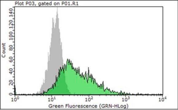
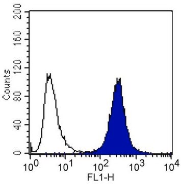
Flow cytometry

Blocks adhesion of cells to extracellular matrix proteins

Induces aggregation of certain lymphoid cells

Does not bind reduced beta.1 integrin on Western blot

Optimal working dilutions must be determined by end user.
Research Category
Cell Structure
Research Sub Category
Integrins

Target description

88 kDa

Physical form

Format: Purified
Protein A Purified mouse immunoglobulin in PBS, no preservatives.
Protein A purified

Storage and Stability

Maintain 6 months at 4°C, from date of shipment. Do not freeze. For maximum recovery of product, centrifuge the original vial after thawing and prior to removing the cap.

Analysis Note

Control
Tonsil, human skin tissue

Other Notes

Concentration: Please refer to the Certificate of Analysis for the lot-specific concentration.

Legal Information

CHEMICON is a registered trademark of Merck KGaA, Darmstadt, Germany

Disclaimer

Unless otherwise stated in our catalog or other company documentation accompanying the product(s), our products are intended for research use only and are not to be used for any other purpose, which includes but is not limited to, unauthorized commercial uses, in vitro diagnostic uses, ex vivo or in vivo therapeutic uses or any type of consumption or application to humans or animals.

Not finding the right product?  

Try our Product Selector Tool.

Storage Class Code

12 - Non Combustible Liquids

WGK

WGK 2

Flash Point(F)

Not applicable

Flash Point(C)

Not applicable


Certificates of Analysis (COA)

Search for Certificates of Analysis (COA) by entering the products Lot/Batch Number. Lot and Batch Numbers can be found on a product’s label following the words ‘Lot’ or ‘Batch’.

Already Own This Product?

Find documentation for the products that you have recently purchased in the Document Library.

Visit the Document Library

Francois Lehembre et al.
The EMBO journal, 27(19), 2603-2615 (2008-09-06)
Loss of expression of the cell-cell adhesion molecule E-cadherin is a hallmark of epithelial-mesenchymal transition (EMT) in development and in the progression from epithelial tumours to invasive and metastatic cancers. Here, we demonstrate that the loss of E-cadherin function upregulates
Simone Bergmann et al.
Journal of cell science, 122(Pt 2), 256-267 (2009-01-02)
By interacting with components of the human host, including extracellular matrix (ECM) proteins, Streptococcus pneumoniae has evolved various strategies for colonization. Here, we characterized the interaction of pneumococci with the adhesive glycoprotein vitronectin and the contribution of this protein to
Leah C Abraham et al.
Experimental cell research, 313(5), 1045-1055 (2007-02-06)
The biodegradation of collagen and the deposition of new collagen-based extracellular matrices are of central importance in tissue remodeling and function. Similarly, for collagen-based biomaterials used in tissue engineering, the degradation of collagen scaffolds with accompanying cellular infiltration and generation
Charles Saby et al.
Oncotarget, 7(18), 24908-24927 (2016-04-29)
Tumor cells are confronted to a type I collagen rich environment which regulates cell proliferation and invasion. Biological aging has been associated with structural changes of type I collagen. Here, we address the effect of collagen aging on cell proliferation
Edwin C K Heng et al.
Journal of cellular biochemistry, 98(2), 409-420 (2006-01-28)
CCN2, (connective tissue growth factor, CTGF) is a matricellular factor associated with fibrosis that plays an important role in the production and maintenance of fibrotic lesions. Increased collagen deposition and accumulation is a common feature of fibrotic tissues. The mechanisms

Our team of scientists has experience in all areas of research including Life Science, Material Science, Chemical Synthesis, Chromatography, Analytical and many others.

Contact Technical Service