AB5896
Anti-P2X3 Receptor Antibody, pain
serum, Chemicon®
Synonym(s):
P2X3 Antibody
Sign Into View Organizational & Contract Pricing
All Photos(1)
About This Item
UNSPSC Code:
12352203
eCl@ss:
32160702
NACRES:
NA.41
Recommended Products
biological source
guinea pig
Quality Level
antibody form
serum
antibody product type
primary antibodies
clone
polyclonal
species reactivity
human, rat, mouse
manufacturer/tradename
Chemicon®
technique(s)
immunocytochemistry: suitable
immunohistochemistry: suitable
NCBI accession no.
UniProt accession no.
shipped in
dry ice
target post-translational modification
unmodified
Gene Information
human ... P2RX3(5024)
Specificity
P2X3 Receptor.
Immunogen
A 15 amino acid peptide corresponding to amino acids 383-397 from the carboxy-terminus of the rat P2X3 Receptor protein.
Control Peptide: Catalog Number AG356
Control Peptide: Catalog Number AG356
Application
Immunohistochemistry: 1:1,000.
Immunocytochemistry: 1:1,000.
Optimal working dilutions must be determined by end user.
APPLICATION NOTES FOR AB5896
IMMUNOHISTOCHEMISTRY
Male Sprague-Dawley rats (b.wt. 100-150g) were anesthetized with sodium pentobarbital and perfused via the ascending aorta with: 1) 50 mL of Ca2+-free Tyrode+s solution followed by 2) a formalin-picric acid fixative (4% paraformaldehyde with 0.4% picric acid in 0.16 M phosphate buffer, pH 6.9) and 3) 10% sucrose in PBS as a cryo-protectant. Tissues were rapidly dissected out and stored overnight in 0.1 M phosphate buffer (pH 7.4) containing 10% sucrose. Slide-mounted tissue sections were incubated with blocking buffer for 1 hour at room temperature. Primary antibody was diluted in blocking buffer to the appropriate working dilution. Blocking buffer was removed and the slides were then incubated at 2-8°C for 18-24 hours with AB5896 (1:1,000). After rinsing in PBS 3 times sections were incubated for 60 minutes at room temperature with Cy3-conjugated secondary antibodies. After mounting in a mixture of PBS and glycerol (1:3) containing 0.1% p-phenylenediamine, sections were examined with a Nikon Microphot-SA epifluorescence microscope.
IMMUNOCYTOCHEMISTRYP
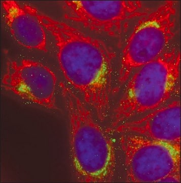
2X3 transfected cells were processed for indirect immunofluorescence. Media was removed and cells were gently washed 3 times with serum-free media. Following fixation procedure, cells were processed for indirect immunofluorescence as above.
Research Category
Neuroscience
Neuroscience
Research Sub Category
Neurotransmitters & Receptors
Neuroinflammation & Pain
Neurotransmitters & Receptors
Neuroinflammation & Pain
This Anti-P2X3 Receptor Antibody, pain is validated for use in IC, IH for the detection of P2X3 Receptor.
Physical form
Serum. Liquid. Contains 0.05% sodium azide.
Storage and Stability
Maintain at -20°C in undiluted aliquots for up to 6 months. Avoid repeated freeze/thaw cycles.
Legal Information
CHEMICON is a registered trademark of Merck KGaA, Darmstadt, Germany
Disclaimer
Unless otherwise stated in our catalog or other company documentation accompanying the product(s), our products are intended for research use only and are not to be used for any other purpose, which includes but is not limited to, unauthorized commercial uses, in vitro diagnostic uses, ex vivo or in vivo therapeutic uses or any type of consumption or application to humans or animals.
Not finding the right product?
Try our Product Selector Tool.
Storage Class Code
10 - Combustible liquids
WGK
WGK 1
Certificates of Analysis (COA)
Search for Certificates of Analysis (COA) by entering the products Lot/Batch Number. Lot and Batch Numbers can be found on a product’s label following the words ‘Lot’ or ‘Batch’.
Already Own This Product?
Find documentation for the products that you have recently purchased in the Document Library.
Juliana Maia Teixeira et al.
Molecular neurobiology, 54(8), 6174-6186 (2016-10-07)
Osteoarthritis (OA) is a degenerative and progressive disease characterized by cartilage breakdown and by synovial membrane inflammation, which results in disability, joint swelling, and pain. The purinergic P2X3 and P2X2/3 receptors contribute to development of inflammatory hyperalgesia, participate in arthritis
Z-J Wang et al.
Anatomy and embryology, 207(4-5), 363-371 (2003-11-19)
Intraganglionic laminar endings (IGLEs) represent the most prominent vagal afferent terminal structures throughout the gastrointestinal tract. They are most prominent in the esophagus and stomach, but can be found down to the distal colon. Their role as mechanosensors as proposed
Zhiyong Chen et al.
Cells, 11(15) (2022-07-28)
The purinergic system plays an important role in pain transmission. Recent studies have suggested that activation of P2-purinergic receptors (P2Rs) may be involved in neuron-satellite glial cell (SGC) interactions in the dorsal root ganglia (DRG), but the details remain unclear.
J L Saloman et al.
Neuroscience, 232, 226-238 (2012-12-04)
Musculoskeletal pain conditions, particularly those associated with temporomandibular disorders (TMD) affect a large percentage of the population. Identifying mechanisms underlying hyperalgesia could contribute to the development of new treatment strategies for the management of TMD and other muscle pain conditions.
Iwan Jones et al.
Scientific reports, 8(1), 15961-15961 (2018-10-31)
The ability to discriminate between diverse types of sensation is mediated by heterogeneous populations of peripheral sensory neurons. Human peripheral sensory neurons are inaccessible for research and efforts to study their development and disease have been hampered by the availability
Our team of scientists has experience in all areas of research including Life Science, Material Science, Chemical Synthesis, Chromatography, Analytical and many others.
Contact Technical Service


