Saltar al contenido
Merck
Todas las fotos(2)

Documentos clave

MABN2719

Sigma-Aldrich

Anti-Neurofilament M/NEFM Antibody, clone 2H3

Sinónimos:

160 kDa neurofilament protein, NF-M, Neurofilament 3, Neurofilament medium polypeptide, Neurofilament triplet M protein

Iniciar sesiónpara Ver la Fijación de precios por contrato y de la organización


About This Item

UNSPSC Code:
12352203
NACRES:
NA.41

biological source

mouse

Quality Level

antibody form

purified antibody

antibody product type

primary antibodies

clone

2H3, monoclonal

mol wt

calculated mol wt 96 kDa
observed mol wt ~160 kDa

purified by

using protein G

species reactivity

rat, human, mouse

packaging

antibody small pack of 100

technique(s)

immunocytochemistry: suitable
immunofluorescence: suitable
immunohistochemistry: suitable
western blot: suitable

isotype

IgG1κ

epitope sequence

Unknown

Protein ID accession no.

UniProt accession no.

storage temp.

2-8°C

Gene Information

rat ... Nefm(24588)

Specificity

Clone 2H3 is a mouse monoclonal antibody that detects Neurofilament-M (NEFM)

Immunogen

Membrane preparations from E14-E15 rat brain tissue.

Application

Quality Control Testing

Evaluated by Western Blotting in Rat brain tissue extracts.

Western Blotting Analysis: A 1:500 dilution of this antibody detected Neurofilament medium polypeptide (NF-M) in Rat brain tissue extract.

Tested Applications

Western Blotting Analysis: A 1:500 dilution from a representative lot detected Neurofilament medium polypeptide (NF-M) in mouse brain tissue extract.

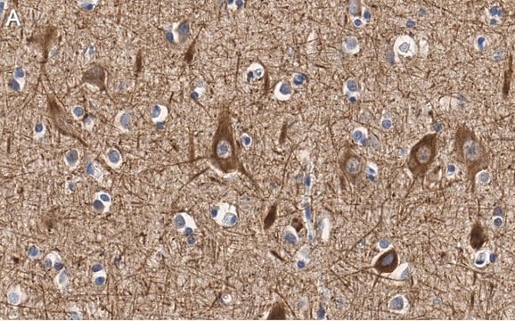
Immunohistochemistry Applications: A representative lot detected Neurofilament medium polypeptide (NF-M) in Immunohistochemistry applications (Clugston, R.D., et al. (2010). Am J Respir Cell Mol Biol. 42(3):276-85; Lysakowski, A., et al. (2011). J Neurosci. 31(27):10101-14; Kridsada, K., et al. (2018). Cell Rep. 23(10):2928-2941; Latremoliere, A., et al. (2018). Cell Rep. 24(7):1865-1879.e.9).

Immunocytochemistry Analysis: A representative lot detected Neurofilament medium polypeptide (NF-M) in Immunocytochemistry applications (Latremoliere, A., et al. (2018). Cell Rep. 24(7):1865-1879.e.9).

Immunofluorescence Analysis: A representative lot detected Neurofilament medium polypeptide (NF-M) in Immunofluorescence applications (Latremoliere, A., et al. (2018). Cell Rep. 24(7):1865-1879.e.9).

Western Blotting Analysis: A representative lot detected Neurofilament medium polypeptide (NF-M) in Western Blotting applications (Fernandez-Cerado, C., et al. (2021). J Neural Transm (Vienna). 128(4):575-587).

Note: Actual optimal working dilutions must be determined by end user as specimens, and experimental conditions may vary with the end user.

Target description

Neurofilament medium polypeptide (UniProt: P12839; also known as NF-M, 160 kDa neurofilament protein, Neurofilament 3, Neurofilament triplet M protein) is encoded by the Nefm (also known as Nef3, Nfm) gene (Gene ID: 24588) in rat. Neurofilaments are intermediate filaments that serve as major elements of the cytoskeleton supporting the axon cytoplasm. They are the most abundant fibrillar components of the axon, being on average 3-10 times more frequent than axonal microtubules. They are built from three intertwined protofibrils, which themselves are composed of two tetrameric protofilament complexes of monomeric proteins. The neurofilament triplet proteins (68/70, 160, and 200 kDa) occur in both the central and peripheral nervous system and are usually neuron specific. The 68/70 kDa NF-L protein can self-assemble into a filamentous structure, however the 160 kDa NF-M and 200 kDa NF-H proteins require the presence of the 68/70 kDa NF-L protein to co-assemble. Neurofilaments contain a number of repeats of the tripeptide KSP (Lys-Ser-Pro) and NF-M is phosphorylated on a number of the serine residues in this motif by protein kinase A and C. Phosphorylation of NF-M results in the formation of interfilament cross bridges that are important in the maintenance of axonal caliber. Deletion of the phosphorylated tail domain of NF-M is reported to inhibit radial growth of axons and reduce their conduction velocities. However, mice expressing NF-M subunits that lack phosphorylation at KSP sites along the tail domain have normal axon calibers and conduction velocities, indicating that the NF-M tail domain, but not tail phosphorylation is crucial for axon radial growth. The level of phosphorylation is shown to be altered developmentally and coincides with a change in the neurofilament function. Mutations in the rod domain region of NF-M have been identified in early onset Parkinson s disease. Also, in the brains of Alzheimer s disease patients, a reciprocal relationship between O-GlcNAcylation and phosphorylation of NF-M has been reported where reduced O-GlcNAcylation is accompanied with increased KSP phosphorylation in NF-M. (Ref.: Yuan, A., et al. (2017). Cold Spring Harb. Perspect. Biol. 9(4); a018309; Yuan, A., et al. (2012). J. Cell Sci. 125(14); 3257-3263).

Physical form

Purified mouse monoclonal antibody IgG1 in buffer containing 0.1 M Tris-Glycine (pH 7.4), 150 mM NaCl with 0.05% sodium azide.

Reconstitution

0.5 mg/mL. Please refer to guidance on suggested starting dilutions and/or titers per application and sample type.

Storage and Stability

Recommended storage: +2°C to +8°C.

Other Notes

Concentration: Please refer to the Certificate of Analysis for the lot-specific concentration.

Disclaimer

Unless otherwise stated in our catalog or other company documentation accompanying the product(s), our products are intended for research use only and are not to be used for any other purpose, which includes but is not limited to, unauthorized commercial uses, in vitro diagnostic uses, ex vivo or in vivo therapeutic uses or any type of consumption or application to humans or animals.

¿No encuentra el producto adecuado?  

Pruebe nuestro Herramienta de selección de productos.

Storage Class

12 - Non Combustible Liquids

wgk_germany

WGK 1

flash_point_f

Not applicable

flash_point_c

Not applicable


Certificados de análisis (COA)

Busque Certificados de análisis (COA) introduciendo el número de lote del producto. Los números de lote se encuentran en la etiqueta del producto después de las palabras «Lot» o «Batch»

¿Ya tiene este producto?

Encuentre la documentación para los productos que ha comprado recientemente en la Biblioteca de documentos.

Visite la Librería de documentos

Nuestro equipo de científicos tiene experiencia en todas las áreas de investigación: Ciencias de la vida, Ciencia de los materiales, Síntesis química, Cromatografía, Analítica y muchas otras.

Póngase en contacto con el Servicio técnico