Saltar al contenido
Merck
Todas las fotos(1)

Documentos clave

MAB343-C

Sigma-Aldrich

Anti-Amyloid Precursor Protein (APP) Antibody

mouse monoclonal, 2.F2.19B4

Sinónimos:

Amyloid beta A4 protein, ABPP, Alzheimer disease amyloid protein, Amyloid precursor protein, APP, APPI, Cerebral vascular amyloid peptide, CVAP, PN-II, PreA4, Protease nexin-II

Iniciar sesiónpara Ver la Fijación de precios por contrato y de la organización


About This Item

Código UNSPSC:
12352203
eCl@ss:
32160702
NACRES:
NA.41

Nombre del producto

Anti-APP Antibody, CT, clone 2.F2.19B4, Ascites Free, clone 2.F2.19B4, from mouse

origen biológico

mouse

Nivel de calidad

forma del anticuerpo

purified immunoglobulin

tipo de anticuerpo

primary antibodies

clon

2.F2.19B4, monoclonal

reactividad de especies

human, mouse

técnicas

affinity binding assay: suitable
immunocytochemistry: suitable
immunofluorescence: suitable
immunohistochemistry: suitable (paraffin)
immunoprecipitation (IP): suitable
western blot: suitable

isotipo

IgG1κ

Nº de acceso NCBI

Nº de acceso UniProt

Condiciones de envío

wet ice

modificación del objetivo postraduccional

unmodified

Información sobre el gen

human ... APP(351)

Descripción general

Amyloid beta A4 protein (UniProt P05067; also known as ABPP, Alzheimer disease amyloid protein, Amyloid precursor protein, APP, APPI, Cerebral vascular amyloid peptide, CVAP, PN-II, PreA4, Protease nexin-II) is encoded by the APP (also known as A4, AD1) gene (Gene ID 351) in human. Amyloid precursor protein (APP) is initially produced with a signal peptide sequence (a.a. 1-17), the removal of which yields the mature protein with a large extracellular portion (a.a. 18-699), followed by a transmembrane segment (a.a. 700-723) and a cytoplasmic (a.a. 724-770) tail. APP can be further processed by the α-, β-, and γ-secretases in two alternative processing pathways. In the non-amyloidogenic pathway, APP is first cleaved by the plasma membrane-localized α-secretase to generate an N-terminal extracellular sAPPα fragment (a.a. 18-687) and a membrane-bound C-terminal fragment C83 (CTFα), which can be further cleaved by γ-secretase to produce a non-toxic small peptide p3 and a cytoplasmic APP intracellular domain (AICD). In the amyloidogenic pathway, APP undergoes β-cleavage in BACE-1 (β-site APP-cleaving enzyme)-enriched endosomes to generate an N-terminal extracellular sAPPβ fragment (a.a. 18-671) and a membrane-bound C-terminal fragment C99 (CTFβ). Subsequent cleavage of C99 by γ-secretase releases the amyloid β peptides, Aβ1-42 (672-713) & Aβ1-40 (672-711), and AICD. Aβ accumulation in the cortical and hippocampal regions of the brain is a major pathological feature of Alzheimer′s disease (AD). Aβ Ser8 phosphorylation is shown to promote Aβ aggregation into oligomeric and fibrillar assemblies and to prevent Aβ proteolytic clearance by certain proteases.

Especificidad

Clone 2.F2.19B4, also known as the Jonas antibody, regcognizes a C-terminal epitope present in 10 of the 11 human spliced isoforms reported by UniProt (P05067), with APP305 being the only spliced isoform lacking the targeted epitope. Clone 2.F2.19B4 recognizes full-length APP, the APP C-terminal fragments (CTFs) produced by α- and β-secretase cleavage, and the APP intracellular domain (AICD) generated by γ-secretase cleavage, but not the sAPPα or sAPPβ fragment. Epitope has been mapped to APP770 a.a. 732-751, which is equivalent to APP695 a.a. 657-676 (Van Vickle, G.D., et al. (2007). Biochemistry. 46(36):10317-10327).

Inmunógeno

APP C-terminal 53-a.a. fragment.
Epitope: APP770 a.a. 732-751 (APP695 a.a. 657-676).

Aplicación

Immunocytochemistry Analysis: A representative lot detected a greatly enhanced nuclear APP immunoreactivity due to an upregulated AICD generation and nuclear localization in HSV-1-infected SH-SY5Y cells by fluorescent immunocytochemistry (De Chiara, G., et al. (2010). PLoS One. 5(11):e13989).
Immunocytochemistry Analysis: A representative lot immunostained neural progenitor cells (NPCs) isolated from the lateral ventricles of E14 mouse brain by fluorescent immunocytochemistry (Ma, Q.H., et al. (2008). Nat. Cell Biol. 10(3):283-294).
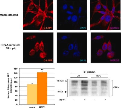
Immunoprecipitation Analysis: A representative lot immunoprecipitated APP and cleaved APP C-terminal fragments from uninfected and HSV-1-infected SH-SY5Y cells (De Chiara, G., et al. (2010). PLoS One. 5(11):e13989).
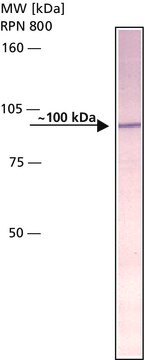
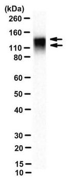
Western Blotting Analysis: A representative lot detected APP and cleaved APP C-terminal fragments in lysates and APP immunoprecipitates from uninfected and HSV-1-infected SH-SY5Y cells (De Chiara, G., et al. (2010). PLoS One. 5(11):e13989).
Western Blotting Analysis: Representative lots detected the stably expressed full-length human APP695 in HEK293-derived BA-3 cells (Hwang, E.M., et al. (2008). Bioorg. Med. Chem. 16(14):6669-6674; Yeon, S.W., et al. (2007). Peptides. 28(4):838-844).
Immunofluorescence Analysis: A representative lot immunostained the walls of the lateral ventricles in E14 mouse brain tissue sections by fluorescent immunohistochemistry (Ma, Q.H., et al. (2008). Nat. Cell Biol. 10(3):283-294).
Affinity Binding Assay Analysis: A representative lot captured the synthetic peptide representing APP770 a.a. 732-751 or APP695 a.a. 657-676 (Van Vickle, G.D., et al. (2007). Biochemistry. 46(36):10317-10327).
Research Category
Neuroscience
Research Sub Category
Neurodegenerative Diseases
This Anti-APP Antibody, CT, clone 2.F2.19B4, Ascites Free is validated for use in Immunohistochemistry (Paraffin), Immunocytochemistry, Immunoprecipitation, Western Blotting, Immunofluorescence, Affinity Binding Assay for the detection of APP.

Calidad

Evaluated by Immunohistochemistry in normal and AD human brain tissue sections.

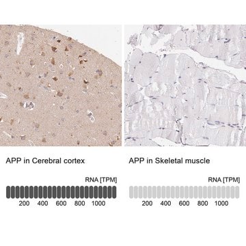
Immunohistochemistry Analysis: A 1:50 dilution from a representative lot detected APP immunoreactivity in normal human cerebral cortex and Alzheimer′s diseased (AD) brain tissue sections.

Descripción de destino

85.18/86.94 kDa (APP770), 75.00/76.76 kDa (L-APP677), 76.90/78.66 kDa (APP695), 77.10/78.87 kDa (L-APP696), 79.01/80.77 kDa (APP714), 81.15/82.92 kDa (L-APP733), 83.06/84.82 kDa (APP751), 83.28/85.04 kDa (L-APP752), 70.79/72.55 kDa (APP639), and 82.76/84.52 kDa (isoform 11) calcualted (full-length mature/pro-form).

Forma física

Format: Purified
Protein G purified.
Purified mouse monoclonal IgG1κ antibody in buffer containing 0.1 M Tris-Glycine (pH 7.4), 150 mM NaCl with 0.05% sodium azide.

Almacenamiento y estabilidad

Stable for 1 year at 2-8°C from date of receipt.

Otras notas

Concentration: Please refer to lot specific datasheet.

Cláusula de descargo de responsabilidad

Unless otherwise stated in our catalog or other company documentation accompanying the product(s), our products are intended for research use only and are not to be used for any other purpose, which includes but is not limited to, unauthorized commercial uses, in vitro diagnostic uses, ex vivo or in vivo therapeutic uses or any type of consumption or application to humans or animals.

¿No encuentra el producto adecuado?  

Pruebe nuestro Herramienta de selección de productos.

Opcional

Referencia del producto
Descripción
Precios

Código de clase de almacenamiento

12 - Non Combustible Liquids

Clase de riesgo para el agua (WGK)

WGK 1

Punto de inflamabilidad (°F)

Not applicable

Punto de inflamabilidad (°C)

Not applicable


Certificados de análisis (COA)

Busque Certificados de análisis (COA) introduciendo el número de lote del producto. Los números de lote se encuentran en la etiqueta del producto después de las palabras «Lot» o «Batch»

¿Ya tiene este producto?

Encuentre la documentación para los productos que ha comprado recientemente en la Biblioteca de documentos.

Visite la Librería de documentos

Nuestro equipo de científicos tiene experiencia en todas las áreas de investigación: Ciencias de la vida, Ciencia de los materiales, Síntesis química, Cromatografía, Analítica y muchas otras.

Póngase en contacto con el Servicio técnico