Saltar al contenido
Merck
Todas las fotos(2)

Documentos clave

MABS2067

Sigma-Aldrich

Anti-HSD3B2 Antibody, clone 6

clone 6, from mouse

Sinónimos:

3-beta-hydroxysteroid dehydrogenase/Delta 5-->4-isomerase type 2, 3 beta-hydroxysteroid dehydrogenase/Delta 5-->4-isomerase type II, 3-beta-HSD II, 3-beta-HSD adrenal and gonadal type

Iniciar sesiónpara Ver la Fijación de precios por contrato y de la organización


About This Item

UNSPSC Code:
12352203
eCl@ss:
32160702
NACRES:
NA.41

biological source

mouse

antibody form

purified immunoglobulin

antibody product type

primary antibodies

clone

6, monoclonal

species reactivity

human

packaging

antibody small pack of 25 μg

technique(s)

ELISA: suitable
immunohistochemistry: suitable (paraffin)
western blot: suitable

isotype

IgG2bκ

NCBI accession no.

UniProt accession no.

target post-translational modification

unmodified

Gene Information

human ... HSD3B2(3284)

General description

3-beta-hydroxysteroid dehydrogenase/Delta 5→4-isomerase type 2 (UniProt: P26439; also known as 3 beta-hydroxysteroid dehydrogenase/Delta 5→4-isomerase type II, 3-beta-HSD II, 3-beta-HSD adrenal and gonadal type) is encoded by the HSD3B2 (also known as HSDB3B) gene (Gene ID: 3284) in human. (3-beta-hydroxysteroid dehydrogenase/Delta 5→4-isomerase type 2 (UniProt: P26439; also known as 3 beta-hydroxysteroid dehydrogenase/Delta 5→4-isomerase type II, 3-beta-HSD II, 3-beta-HSD adrenal and gonadal type) is encoded by the HSD3B2 (also known as HSDB3B) gene (Gene ID: 3284) in human. Humans have two HSD3B isozymes. HSD3B1 is mainly expressed in the placenta, mammary and, prostate glands, and other peripheral tissues including the skin and adipose tissue and HSD3B2 is expressed primarily in the adrenal and gonads. They exhibit about 93% sequence homology. 3-beta-HSD II is a single-pass membrane protein found on mitochondrial and endoplasmic reticulum membranes. It is expressed in adrenal gland, testis and ovary. It is a bifunctional enzyme that catalyzes the oxidative conversion of Delta 5-ene-3-beta-hydroxy steroid and the oxidative conversion of ketosteroids. It plays a crucial role in the biosynthesis of all classes of hormonal steroids. Mutations in HSD3B2 gene are reported to cause adrenal hyperplasia that is characterized by androgen excess, rapid somatic growth during childhood in both sexes with premature closure of the epiphyses and short adult stature. Mild HSD3B2 deficiency in hyperandrogenic females is associated with characteristic traits of polycystic ovary syndrome, such as insulin resistance and luteinizing hormone hypersecretion (Ref.: Gomez-Sanchez, CE et al. (2017). Steroids 127; 56-61).

Specificity

Clone 6 specifically detects HSDB32 in human tissue. it targets an epitope with in 14 amino acids from the N-terminal region.

Immunogen

KLH-conjugated linear peptide corresponding to 14 amino acids from the N-terminal region of human 3-beta-hydroxysteroid dehydrogenase/Delta 5→4-isomerase type 2 (3-beta-HSD).

Application

Anti-HSD3B2, clone 6, Cat. No. MABS2067, is a mouse monoclonal antibody that detects 3-beta-HSD II (HSD3B2) and has been tested for use in ELISA, Immunohistochemistry (Paraffin), and Western Blotting.
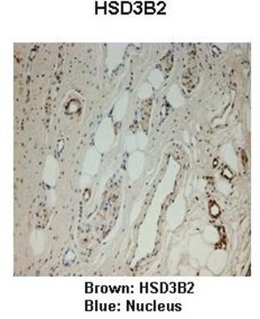
Immunohistochemistry (Paraffin) Analysis: A representative lot detected HSD3B2 in Immunohistochemistry appliations (Gomez-Sanchez, C.E., et al. (2017). Steroids. 127:56-61).

Western Blotting Analysis: Analysis: A representative lot detected HSD3B2 in Western Blotting appliations (Gomez-Sanchez, C.E., et al. (2017). Steroids. 127:56-61).

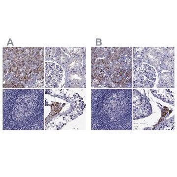
Immunohistochemistry (Paraffin) Analysis: A 1:50-250 dilution from a representative lot detected HSD3B2 in human adrenal gland and human placenta tissue sections.

ELISA Analysis: A representative lot detected HSD3B2 in ELISA appliations (Gomez-Sanchez, C.E., et al. (2017). Steroids. 127:56-61).
Research Category
Signaling

Quality

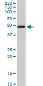
Evaluated by Western Blotting in human adrenal gland tissue lysate.

Western Blotting Analysis: 1 µg/mL of this antibody detected HSD3B2 in human adrenal gland tissue lysate.

Target description

~40 kDa observed; 42.05 kDa calculated. Uncharacterized bands may be observed in some lysate(s).

Physical form

Format: Purified
Protein G purified
Purified mouse monoclonal antibody IgG2b in buffer containing 0.1 M Tris-Glycine (pH 7.4), 150 mM NaCl with 0.05% sodium azide.

Storage and Stability

Stable for 1 year at 2-8°C from date of receipt.

Other Notes

Concentration: Please refer to lot specific datasheet.

Disclaimer

Unless otherwise stated in our catalog or other company documentation accompanying the product(s), our products are intended for research use only and are not to be used for any other purpose, which includes but is not limited to, unauthorized commercial uses, in vitro diagnostic uses, ex vivo or in vivo therapeutic uses or any type of consumption or application to humans or animals.

¿No encuentra el producto adecuado?  

Pruebe nuestro Herramienta de selección de productos.


Certificados de análisis (COA)

Busque Certificados de análisis (COA) introduciendo el número de lote del producto. Los números de lote se encuentran en la etiqueta del producto después de las palabras «Lot» o «Batch»

¿Ya tiene este producto?

Encuentre la documentación para los productos que ha comprado recientemente en la Biblioteca de documentos.

Visite la Librería de documentos

Nuestro equipo de científicos tiene experiencia en todas las áreas de investigación: Ciencias de la vida, Ciencia de los materiales, Síntesis química, Cromatografía, Analítica y muchas otras.

Póngase en contacto con el Servicio técnico