Saltar al contenido
Merck
Todas las fotos(1)

Documentos clave

MABF2820

Sigma-Aldrich

Anti-HCV E2 Antibody, clone AP33

Sinónimos:

HCV envelope glycoprotein E2

Iniciar sesiónpara Ver la Fijación de precios por contrato y de la organización


About This Item

UNSPSC Code:
12352203
NACRES:
NA.41

biological source

mouse

Quality Level

antibody form

purified antibody

antibody product type

primary antibodies

clone

AP33, monoclonal

mol wt

observed mol wt ~60 kDa

purified by

using protein G

species reactivity

virus

packaging

antibody small pack of 100 μg

technique(s)

ELISA: suitable
electron microscopy: suitable
immunoprecipitation (IP): suitable
inhibition assay: suitable
neutralization: suitable
western blot: suitable

isotype

IgG1κ

epitope sequence

N-terminal half

Protein ID accession no.

UniProt accession no.

storage temp.

-10 to -25°C

target post-translational modification

unmodified

Gene Information

vaccinia virus ... E2(684)

General description

Genome polyprotein (UniProt: P27958) is encoded by the POLY gene (Gene ID: 951475) in Hepatitis C virus. HCV is a small (~55-65 nm), enveloped, positive-sense single-stranded RNA virus that is a causative factor for hepatitis C and hepatocellular carcinoma. HCV expresses several proteins that promote viral replication. Genome polyprotein of HCV can be cleaved into 11 different chains. The mature core protein packages viral RNA to form a viral nucleocapsid and promotes virion budding. It also prevents the establishment of cellular antiviral state by blocking the interferon- /β and interferon- signaling pathways and by inducing STAT1 degradation. Envelope proteins E1 and E2 that form a heterodimer are heavily glycosylated and are involved in virus attachment to the host cell, virion internalization through clathrin-dependent endocytosis and fusion with host membrane. In HCV, Viroprotein P7, a heptameric ion channel protein, plays an essential role in the assembly, envelopment, and secretion of viral particles. It also contains NS2, a cysteine protease and NS3, a serine protease. NS2 is required for the proteolytic auto-cleavage between NS2 and NS3. NS3 binds a single ATP and catalyzes the unzipping of a single base pair of dsRNA. Non-structural protein 4A (NS4A), a peptide cofactor, forms a non-covalent complex with the N-terminal of NS3 and this complex prevents phosphorylation of host IRF3, which also prevents the establishment of dsRNA induced antiviral state. NS4B induces a specific membrane alteration that serves as a scaffold for the virus replication complex. Non-structural protein 5A (NS5A), a multi-functional phosphoprotein, is an RNA-binding peripheral membrane protein that is reported to antagonize numerous cellular pathways, including the antiviral interferon-a response. NS5A exists as hypo- and hyper-phosphorylated forms and the dynamic transition between these two states is involved in the functions of NS5A. Hyperphosphorylation occurs primarily at its six serine residues within the low complexity sequence of NS5A. Phosphorylated NS5A is shown to be essential for viral replication and assembly. Clone AP33 recognizes an epitope within the N-terminal portion of the E2 ectodomain (aa 412 to 423). It is shown to inhibit the interaction of truncated E2 (E2(660)), full-length E1E2 complex expressed in mammalian cells, and of HCV virus-like particles with CD81. It also displays potent neutralizing activity against HCV carrying E1E2 of all genotypes. (Ref.: Owsianka, A., et al. (2005). J. Virol. 79(17); 11095-11104; Clayton, RF., et al. (2002). J. Virol. 76(15); 7672-7682; Owsianka, A., et al. (2001). J. Gen. Virol. 82(8); 1877-1883).

Specificity

Clone AP33 is a mouse monoclonal antibody that detects envelope glycoprotein E2. It targets an epitope within 12 amino acids from the N-terminal half.

Immunogen

His-tagged recombinant form of mammalian cell-expressed HCV strain Gla E1E2 emulsified in Freund′s complete adjuvant.

Application

Quality Control Testing

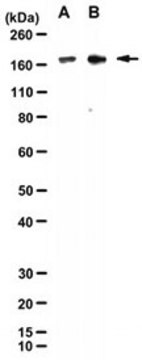
Evaluated by Western Blotting in lysate from HEK293T cells transfected with a plasmid expressing the HCV E1 and E2 glycoproteins.

Western Blotting Analysis: A 1:1,000 dilution of this antibody detected HCV E2 in lysate from HEK293T cells transfected with a plasmid expressing the HCV E1 and E2 glycoproteins, but not in lysates from untransfected cells.

Tested Applications

Inhibition: A representative lot Inhibited the interaction of virus-like particles (VLPs) with CD81 and inhibited infection by HCVpp of diverse genotypes.(Owsianka, A., et al. (2001). J Gen Virol. 82(Pt8):1877-1883; Owsianka, A., et al. (2005). J Virol. 79(17):11095-104).

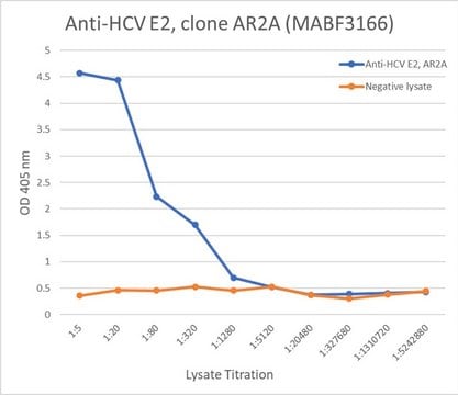
ELISA: A representative lot detected HCV E2 in ELISA application. (Owsianka, A., et al. (2005). J Virol. 79(17):11095-104; Rychlowska, M., et al. (2011). J Gen Virol. 92(Pt10)2249-2261; Vasiliauskaite, I., et al. (2017). mBio. 8(3):e00382-17).

Electron Microscopy: A representative lot detected virus like particles of HCV in Electron Cryomicroscopy application (Owsianka, A., et al. (2001). J Gen Virol. 82(Pt8):1877-1883; Clayton, R.F., et al. (2002). J Virol. 76(15):7672-82).

Western Blotting Analysis: A representative lot detected HCV E2 in Western Blotting application (Clayton, R.F., et al. (2002). J Virol. 76(15):7672-82; Rychlowska, M., et al. (2011). J Gen Virol. 92(Pt10)2249-2261; Hu, Z., et al. (2020). Cell Chem Biol. 27(7):780-792.e5).

Immunoprecipitation Analysis: A representative lot immunoprecipitated HCV E2 in Immunoprecipitation application (Owsianka, A., et al. (2005). J Virol. 79(17):11095-104).

Neutralizing: A representative lot neutralized HCV infection across all major genotypes of HCV (Owsianka, A., et al. (2005). J Virol. 79(17):11095-104; Potter, J.A., et al. (2012). J Virol. 86(23):12923-32).

Note: Actual optimal working dilutions must be determined by end user as specimens, and experimental conditions may vary with the end user.

Physical form

Purified mouse monoclonal antibody IgG1 in PBS without azide.

Reconstitution

1.0 mg/mL. Please refer to guidance on suggested starting dilutions and/or titers per application and sample type.

Storage and Stability

Store at -10°C to -25°C. Handling Recommendations: Upon receipt and prior to removing the cap, centrifuge the vial and gently mix the solution. Aliquot into microcentrifuge tubes and store at -20°C. Avoid repeated freeze/thaw cycles, which may damage IgG and affect product performance.

Other Notes

Concentration: Please refer to the Certificate of Analysis for the lot-specific concentration.

Disclaimer

Unless otherwise stated in our catalog or other company documentation accompanying the product(s), our products are intended for research use only and are not to be used for any other purpose, which includes but is not limited to, unauthorized commercial uses, in vitro diagnostic uses, ex vivo or in vivo therapeutic uses or any type of consumption or application to humans or animals.

¿No encuentra el producto adecuado?  

Pruebe nuestro Herramienta de selección de productos.

Storage Class

12 - Non Combustible Liquids

wgk_germany

WGK 2

flash_point_f

Not applicable

flash_point_c

Not applicable


Certificados de análisis (COA)

Busque Certificados de análisis (COA) introduciendo el número de lote del producto. Los números de lote se encuentran en la etiqueta del producto después de las palabras «Lot» o «Batch»

¿Ya tiene este producto?

Encuentre la documentación para los productos que ha comprado recientemente en la Biblioteca de documentos.

Visite la Librería de documentos

Nuestro equipo de científicos tiene experiencia en todas las áreas de investigación: Ciencias de la vida, Ciencia de los materiales, Síntesis química, Cromatografía, Analítica y muchas otras.

Póngase en contacto con el Servicio técnico