Accéder au contenu
Merck
Toutes les photos(2)

Key Documents

SAB4500362

Sigma-Aldrich

Anti-Collagen I antibody produced in rabbit

affinity isolated antibody

Synonyme(s) :

α 2 type i collagen, co1a2, col1a2, collagen, type i

Se connecterpour consulter vos tarifs contractuels et ceux de votre entreprise/organisme


About This Item

Code UNSPSC :
12352203
Nomenclature NACRES :
NA.41

Source biologique

rabbit

Conjugué

unconjugated

Forme d'anticorps

affinity isolated antibody

Type de produit anticorps

primary antibodies

Clone

polyclonal

Forme

buffered aqueous solution

Poids mol.

antigen 129 kDa

Espèces réactives

mouse, human, rat

Concentration

~1 mg/mL

Technique(s)

ELISA: 1:5000
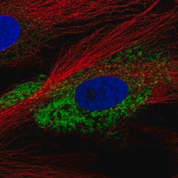
immunofluorescence: 1:100-1:500
immunohistochemistry: 1:50-1:100

Numéro d'accès NCBI

Numéro d'accès UniProt

Conditions d'expédition

wet ice

Température de stockage

−20°C

Modification post-traductionnelle de la cible

unmodified

Informations sur le gène

human ... COL1A2(1278)

Description générale

Collagen I is encoded by the gene COL1A2 (collagen type I alpha 2). The gene is located on human chromosome 7q21.3. COL1A2 is a fibril forming collagen, found in bone, dermis, tendon, ligaments and cornea.
Collagen is the major structural component of extracellular matrices found in connective tissues and internal organs, but is most prevalent in the dermis, tendons, and bones. It is classified into a number of structurally and genetically distinct types. Type I collagen is a heterodimer composed of two α1(I) chains and one α2(I) chain that spontaneously forms a triple helix scaffold at neutral pH and 37 °C. Collagen type I is an excellent substrate for the culture of hepatocytes, fibroblasts, spinal ganglion, muscle cells, Schwann cells, embryonic lung cells, epithelial cells, and a number of other cell lines. It has also been used in the study of growth, differentiation, migration of cell lines, and tissue morphogenesis during development.
Rabbit polyclonal anti-Collagen I, N-Terminal antibody reacts with mouse, rat, and human type I collagens.

Immunogène

The antiserum was produced against synthesized peptide derived from human Collagen I.

Immunogen Range: 1-50

Application

Anti-Collagen I antibody produced in rabbit has been used in western blotting and immunostaining.
Applications in which this antibody has been used successfully, and the associated peer-reviewed papers, are given below.
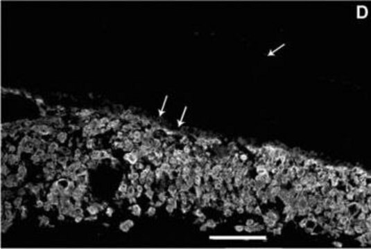
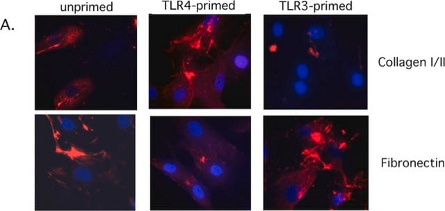
Immunofluorescence (1 paper)
Rabbit polyclonal anti-Collagen I, N-Terminal antibody is used to tag collagen type I for detection and quantitation by immunocytochemical and immunohistochemical (IHC) techniques such as ELISA, immunoblotting, and immunoprecipitation. It is used as a probe to determine the presence and roles of collagen type I as a cell substrate.

Actions biochimiques/physiologiques

Collagen I interacts with cytoskeleton and regulates cell motility. Mutations in this gene is linked to osteoporotic fracture, osteogenesis imperfecta and recessive classical type Ehlers-Danlos syndrome. It is also associated with idiopathic osteoporosis and atypical Marfan syndrome.

Caractéristiques et avantages

Evaluate our antibodies with complete peace of mind. If the antibody does not perform in your application, we will issue a full credit or replacement antibody. Learn more.

Forme physique

Rabbit IgG in phosphate buffered saline (without Mg2+ and Ca2+), pH 7.4, 150mM NaCl, 0.02% sodium azide and 50% glycerol.

Clause de non-responsabilité

Unless otherwise stated in our catalog or other company documentation accompanying the product(s), our products are intended for research use only and are not to be used for any other purpose, which includes but is not limited to, unauthorized commercial uses, in vitro diagnostic uses, ex vivo or in vivo therapeutic uses or any type of consumption or application to humans or animals.

Vous ne trouvez pas le bon produit ?  

Essayez notre Outil de sélection de produits.

Code de la classe de stockage

12 - Non Combustible Liquids

Classe de danger pour l'eau (WGK)

nwg

Point d'éclair (°F)

Not applicable

Point d'éclair (°C)

Not applicable


Certificats d'analyse (COA)

Recherchez un Certificats d'analyse (COA) en saisissant le numéro de lot du produit. Les numéros de lot figurent sur l'étiquette du produit après les mots "Lot" ou "Batch".

Déjà en possession de ce produit ?

Retrouvez la documentation relative aux produits que vous avez récemment achetés dans la Bibliothèque de documents.

Consulter la Bibliothèque de documents

COL1A2 is a Novel Biomarker to Improve Clinical Prediction in Human Gastric Cancer: Integrating Bioinformatics and Meta-Analysis
Rong L, et al.
Pathology Oncology Research, 24(1), 129-134 (2018)
Fine mapping of the autosomal dominant split hand/split foot locus on chromosome 7, band q21. 3-q22. 1
Scherer SW, et al.
American Journal of Human Genetics, 55(1), 12-12 (1994)
Respiratory syncytial virus infection accelerates lung fibrosis through the unfolded protein response in a bleomycin-induced pulmonary fibrosis animal model
Wang L, et al.
Molecular Medicine Reports, 16(1), 310-316 (2017)
Lina Wang et al.
Molecular medicine reports, 16(1), 310-316 (2017-05-13)
Emerging evidence has demonstrated that endoplasmic reticulum stress (ER) is involved in the pathogenesis of idiopathic pulmonary fibrosis, however, the underlying mechanism remains unclear. Viral infection often triggers a hyperinflammatory response by an expansion of the ER. The present study
Nastaran Tarban et al.
Cells, 11(8) (2022-04-24)
Skeletal muscle repair is initiated by local inflammation and involves the engulfment of dead cells (efferocytosis) by infiltrating macrophages at the injury site. Macrophages orchestrate the whole repair program, and efferocytosis is a key event not only for cell clearance

Notre équipe de scientifiques dispose d'une expérience dans tous les secteurs de la recherche, notamment en sciences de la vie, science des matériaux, synthèse chimique, chromatographie, analyse et dans de nombreux autres domaines..

Contacter notre Service technique