Accéder au contenu
Merck
Toutes les photos(3)

Key Documents

MAB16985

Sigma-Aldrich

Anti-MCAM Antibody, clone P1H12

clone P1H12, Chemicon®, from mouse

Synonyme(s) :

CD146 antigen, Cell surface glycoprotein P1H12, Melanoma-associated antigen A32, Melanoma-associated antigen MUC18, S-endo 1 endothelial-associated antigen, melanoma adhesion molecule, melanoma cell adhesion molecule

Se connecterpour consulter vos tarifs contractuels et ceux de votre entreprise/organisme


About This Item

Code UNSPSC :
12352203
eCl@ss :
32160702
Nomenclature NACRES :
NA.41

Source biologique

mouse

Niveau de qualité

Forme d'anticorps

purified immunoglobulin

Type de produit anticorps

primary antibodies

Clone

P1H12, monoclonal

Espèces réactives

mouse, canine, human

Ne doit pas réagir avec

rat

Conditionnement

antibody small pack of 25 μg

Fabricant/nom de marque

Chemicon®

Technique(s)

ELISA: suitable
flow cytometry: suitable
immunocytochemistry: suitable
immunohistochemistry: suitable
immunoprecipitation (IP): suitable
western blot: suitable

Isotype

IgG1

Adéquation

not suitable for immunohistochemistry (Paraffin)

Numéro d'accès NCBI

Numéro d'accès UniProt

Conditions d'expédition

ambient

Température de stockage

2-8°C

Modification post-traductionnelle de la cible

unmodified

Informations sur le gène

human ... MCAM(4162)

Description générale

MUC18 antigen (CD146), a member of the immuoglobulin superfamily, is an intrinsic membrane glycoprotein of 110-120 kDa found on the surface of endothelial cells, bone marrow fibroblasts and various melanomas. MUC18 has been used as a marker of tumor progression in human melanoma because expression in those tumors correlates strongly with poor prognosis and the development of metastic disease. In addition, a number of human T, B and myeloid leukemic cell lines seem to express MUC18. The close structural relationship with N-CAM and related molecules suggests that MUC18 may be also a developmentally regulated cell adhesion molecule (Melanoma adhesion molecule or MCAM).

Spécificité

Detects circulating endothelial cells in human. Expected to cross-react with dog & mouse.

Immunogène

Immunization of mice with HUVEC.

Application

Anti-MCAM Antibody, clone P1H12 detects level of MCAM & has been published & validated for use in ELISA, FC, IC, IH, IP & WB.
Immunocytochemistry:
1-10 μg/mL of a previous lot worked in immunocytochemistry.Works best on EDTA or Trypsin lifted endothelial cells.

Immunohistochemistry:
1-10 µg/mL. 4% PFA for 30min RT or <2hrs at 4°C. Block w/ 1%BSA/0.2% tween20/PBS for 30min. Works well in frozen tissue; fixed or unfixed.

Immunoprecipitation:
1-10 μg/mL of a previous lot worked in immunoprecipitation.

ELISA:
1-10 μg/mL of a previous lot worked in ELISA.

FACS Analysis:
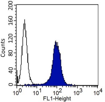
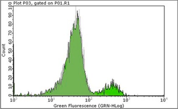
1-10 μg/mL of a previous lot worked in FACS.

Optimal working dilutions must be determined by end user.

Qualité

Routinely evaluated by Western Blot on HUVEC lysates.

Western Blot Analysis:
1:500 dilution of this lot detected MCAM on 10μg of HUVEC lysates.

Description de la cible

110 kDa

Forme physique

Format: Purified
Purified mouse monoclonal IgG1 in buffer containing 0.02 M PB with 0.25 M NaCl, pH=7.6, with 0.1% sodium azide.

Remarque sur l'analyse

Control
HUVEC cells.

Autres remarques

Concentration: Please refer to the Certificate of Analysis for the lot-specific concentration.

Informations légales

CHEMICON is a registered trademark of Merck KGaA, Darmstadt, Germany

Vous ne trouvez pas le bon produit ?  

Essayez notre Outil de sélection de produits.

Code de la classe de stockage

12 - Non Combustible Liquids

Classe de danger pour l'eau (WGK)

WGK 2

Point d'éclair (°F)

Not applicable

Point d'éclair (°C)

Not applicable


Certificats d'analyse (COA)

Recherchez un Certificats d'analyse (COA) en saisissant le numéro de lot du produit. Les numéros de lot figurent sur l'étiquette du produit après les mots "Lot" ou "Batch".

Déjà en possession de ce produit ?

Retrouvez la documentation relative aux produits que vous avez récemment achetés dans la Bibliothèque de documents.

Consulter la Bibliothèque de documents

Keiich Hirono et al.
Journal of the American College of Cardiology, 48(6), 1257-1264 (2006-09-19)
This study investigated patients with acute Kawasaki disease (KD) to validate myeloid-related protein (MRP)-8/MRP-14 as a marker of disease activity and severity of coronary artery lesion development. Both MRP-8 and -14, which are S100-proteins secreted by activated neutrophils and monocytes
Feng Li et al.
Bone, 47(3), 546-555 (2010-06-24)
Currently, there are conflicting data in literature regarding contribution of bone marrow stromal cells (BMSCs) to bone formation when the cells are systemically delivered in recipient animals. To understand if BMSCs contribute to bone cell phenotype and bone formation in
Ivan Depasquale et al.
Cancer cell international, 6, 9-9 (2006-04-01)
Lovastatin and other statins may reduce the development of melanomas. The effects on melanoma cells and their ability to enhance angiogenesis in a co-culture system presented an opportunity to assess whether Lovastatin act on melanoma cells, HUVEC or both types
D Baeten et al.
Annals of the rheumatic diseases, 63(5), 489-493 (2004-04-15)
It has been suggested that the immunopathology of rheumatoid nodules parallels that of inflamed synovium in rheumatoid arthritis (RA). To analyse the effect of infliximab on the immunopathology of rheumatoid nodules in order to provide new insights into the relationship
Catherine Larochelle et al.
Brain : a journal of neurology, 135(Pt 10), 2906-2924 (2012-09-15)
In multiple sclerosis, encephalitogenic CD4(+) lymphocytes require adhesion molecules to accumulate into central nervous system inflammatory lesions. Using proteomic techniques, we identified expression of melanoma cell adhesion molecule (MCAM) on a subset of human effector memory CD4(+) lymphocytes and on

Notre équipe de scientifiques dispose d'une expérience dans tous les secteurs de la recherche, notamment en sciences de la vie, science des matériaux, synthèse chimique, chromatographie, analyse et dans de nombreux autres domaines..

Contacter notre Service technique