Accéder au contenu
MilliporeSigma
Toutes les photos(2)

Documents

MABT339

Sigma-Aldrich

Anti-ZO-1 Antibody, clone 5G6.1

clone 5G6.1, from mouse

Synonyme(s) :

Tight junction protein ZO-1, Tight junction protein 1, Zona occludens protein 1, Zonula occludens protein 1, ZO-1

Se connecterpour consulter vos tarifs contractuels et ceux de votre entreprise/organisme


About This Item

Code UNSPSC :
12352203
eCl@ss :
32160702
Nomenclature NACRES :
NA.41

Source biologique

mouse

Niveau de qualité

Forme d'anticorps

purified immunoglobulin

Type de produit anticorps

primary antibodies

Clone

5G6.1, monoclonal

Espèces réactives

rat, human

Technique(s)

immunohistochemistry: suitable (paraffin)
western blot: suitable

Isotype

IgG2aκ

Numéro d'accès NCBI

Numéro d'accès UniProt

Conditions d'expédition

wet ice

Modification post-traductionnelle de la cible

unmodified

Informations sur le gène

human ... TJP1(7082)

Description générale

Tight junction protein ZO-1 (UniProt Q07157; also known as Tight junction protein 1, Zona occludens protein 1, Zonula occludens protein 1) is encoded by the TJP1 gene (also known as ZO1) (Gene ID 7082) in human. The tight junction (TJ) constitutes the barrier between the apical and the basolateral domains of the plasma membrane. The assembly and permeability of this barrier are dependent on the zonula occludens (ZO) membrane-associated guanylate kinase (MAGUK) proteins ZO-1, ZO-2, and ZO-3. ZO-1, a 210-225 kDa protein, is found at the submembranous domain of TJs in epithelia and endothelia. ZO-1 contains three PDZ domains (PDZ1/aa23-109; PDZ2/aa181-261; PDZ3/aa422-502) at its N-treminal end, followed by an SH3 domain (aa519-580), a GUK/GK homology domain (aa632-782), an acidic domain (aa817-894), an alpha spliced domain (aa921-1000), and a C-terminal proline-rich/PR domain. ZO-1 is a phosphoprotein and a known substrate of serine/threonine kinases ZAK and of PKC. MAPK signaling pathway regulates tyrosine phosphorylation of ZO-1, and MEK1 inhibition in Ras transformed epithelial cells is reported to result in tyrosine phosphorylation of ZO-1 and occludin. ZO-1 interacts with claudins, JAM, ZO-2, and ZO-3 through its PDZ domains, while its GK module mediates interaction with occludin. ZO-1 also binds actin cytoskeleton and actin-binding protein 4.1 through its carboxyl terminal end. In addition, ZO-1 associates with AF-6 and cingulin at the TJ, as well as with the adherens junction protein alpha-catenin and with the gap junction proteins connexins 43 and 45.

Immunogène

GST-tagged recombinant protein corresponding to human ZO-1.

Application

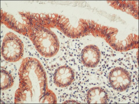
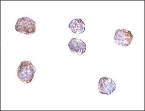
Immunohistochemistry Analysis: A 1:1,000 dilution from a representative lot detected ZO-1 in human and rat kidney tissues.
Research Category
Cell Structure
Research Sub Category
Adhesion (CAMs)
This Anti-ZO-1 Antibody, clone 5G6.1 is validated for use in Western Blotting, Immunocytochemistry, Flow Cytometry, ELISA for the detection of ZO-1 .

Qualité

Evaluated by Western Blotting in HCT116 cell lysate.

Western Blotting Analysis: 0.5 µg/mL of this antibody detected ZO-1 in 200 µg of HCT116 cell lysate.

Description de la cible

~195 kDa observed

Forme physique

Format: Purified
Protein G Purified
Purified mouse monoclonal IgG2aκ antibody in buffer containing 0.1 M Tris-Glycine (pH 7.4), 150 mM NaCl with 0.05% sodium azide.

Stockage et stabilité

Stable for 1 year at 2-8°C from date of receipt.

Autres remarques

Concentration: Please refer to lot specific datasheet.

Clause de non-responsabilité

Unless otherwise stated in our catalog or other company documentation accompanying the product(s), our products are intended for research use only and are not to be used for any other purpose, which includes but is not limited to, unauthorized commercial uses, in vitro diagnostic uses, ex vivo or in vivo therapeutic uses or any type of consumption or application to humans or animals.

Not finding the right product?  

Try our Outil de sélection de produits.

Code de la classe de stockage

12 - Non Combustible Liquids

Classe de danger pour l'eau (WGK)

WGK 1

Point d'éclair (°F)

Not applicable

Point d'éclair (°C)

Not applicable


Certificats d'analyse (COA)

Recherchez un Certificats d'analyse (COA) en saisissant le numéro de lot du produit. Les numéros de lot figurent sur l'étiquette du produit après les mots "Lot" ou "Batch".

Déjà en possession de ce produit ?

Retrouvez la documentation relative aux produits que vous avez récemment achetés dans la Bibliothèque de documents.

Consulter la Bibliothèque de documents

Duobin Zhang et al.
Journal of leukocyte biology, 111(5), 953-966 (2021-09-30)
Ischemic stroke (IS) can cause disability and death, and microglia as the immune component of the CNS can release inflammatory factors and participate in blood-brain barrier (BBB) dysfunction. This study aimed to investigate the effects of long noncoding RNA (lncRNA)
Muhammad Ali et al.
Stem cell reports, 16(9), 2320-2335 (2021-08-07)
Here, we evaluate the efficacy of cryopreserved human embryonic stem cell (hESC)-derived corneal endothelial cells (CECs) to form a functional monolayer of corneal endothelium (CE) in rabbits and monkeys. We injected cryopreserved hESC-derived CECs into the anterior chamber of rabbits
Adam M Jorgensen et al.
Tissue engineering. Part A, 26(9-10), 512-526 (2019-12-22)
Over 1 million burn injuries are treated annually in the United States, and current tissue engineered skin fails to meet the need for full-thickness replacement. Bioprinting technology has allowed fabrication of full-thickness skin and has demonstrated the ability to close
Danielle B Buglak et al.
Angiogenesis, 23(4), 567-575 (2020-07-24)
Proper blood vessel formation requires coordinated changes in endothelial cell polarity and rearrangement of cell-cell junctions to form a functional lumen. One important regulator of cell polarity is the centrosome, which acts as a microtubule organizing center. Excess centrosomes perturb

Articles

Millicell® hanging cell culture inserts for staining and microscopic analysis directly from the insert. Learn to utilize hanging cell culture inserts in barrier measurements.

Contenu apparenté

Step-by-step protocol for generating apical-out human gut organoids for microbiome, ADME/Tox, viral and gastrointestinal related disease research. See the complete organoid culture protocol.

Monitor barrier formation using colon PDOs, iPSC-derived colon organoids, Millicell® cell culture inserts, and the Millicell® ERS. 3.0.

Notre équipe de scientifiques dispose d'une expérience dans tous les secteurs de la recherche, notamment en sciences de la vie, science des matériaux, synthèse chimique, chromatographie, analyse et dans de nombreux autres domaines..

Contacter notre Service technique