Skip to Content
MilliporeSigma
All Photos(3)

Key Documents

ABS1593

Sigma-Aldrich

Anti-sFRP-1 Antibody

from rabbit

Synonym(s):

Secreted frizzled-related protein 1, FRP-1, SARP-2, Secreted apoptosis-related protein 2, sFRP-1

Sign Into View Organizational & Contract Pricing


About This Item

UNSPSC Code:
12352203
eCl@ss:
32160702
NACRES:
NA.41

biological source

rabbit

Quality Level

antibody form

purified antibody

antibody product type

primary antibodies

clone

polyclonal

species reactivity

human, mouse

technique(s)

immunohistochemistry: suitable
neutralization: suitable
western blot: suitable

NCBI accession no.

UniProt accession no.

shipped in

ambient

target post-translational modification

unmodified

Gene Information

human ... SFRP1(6422)

General description

Secreted frizzled-related protein 1 (UniProt Q8N474; also known as FRP-1, SARP-2, Secreted apoptosis-related protein 2, sFRP-1) is encoded by the SFRP1 (also known as FRP, FRP1, SARP2) gene (Gene ID 6422) in human. Canonical Wnt signaling is constitutively suppressed in the absence of Wnt, and its activation in the presence of Wnt can be blocked by secreted inhibitors, including the dickkopf-related proteins (DKK) and the secreted frizzled-related proteins (sFRP1-5). sFRPs share homology with the FZD receptor and compete againt FZD for Wnt binding in the extracellular space, sFRPs may also exert their effects by interacting with each other and FZD receptors. sFRP-1 is present during both cardiac development and adult life and is abundantly expressed in the mouse and human heart. Study conducted with Sfrp1-knockout mice indicates a role of sFRP-1 in age-related cardiac deterioration and fibrosis. Human sFRP-1 is produced with a signal peptide sequence (a.a. 1-31), the removal of which yields the mature protein (a.a. 32-314) with an N-terminal FZ (a.a. 53-169) domain, followed by a netrin (NTR) domain (a.a. 186-306).

Specificity

This polyclonal antibody neutralized the inhibitory activity of sFRP-1 (Scheel, C., et al. (2011). Cell. 145(6):926 940; Häusler, K.D., et al. (2004). J. Bone Miner. Res. 19(11):1873-1881).

Immunogen

Mammalian expressed human sFRP-1, purified from condition culture medium (Häusler, K.D., et al. (2004). J. Bone Miner. Res. 19(11):1873-1881; Uren, A., et al. (2000). J. Biol. Chem. 275(6):4374-4382).

Application

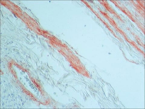
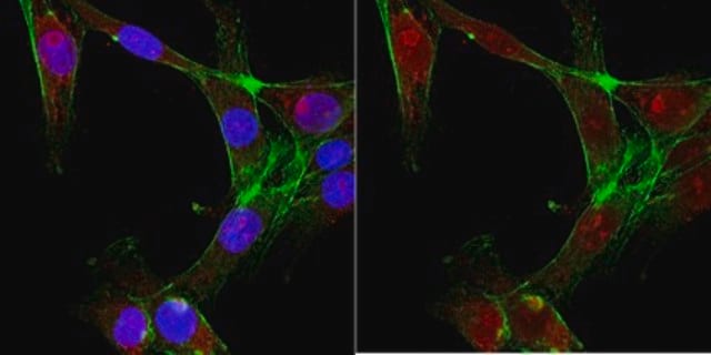
Detect sFRP-1 using this rabbit polyclonal Anti-sFRP-1, Cat. No. ABS1593, validated for use in Immunohistochemistry, Neutralization, and Western Blotting.
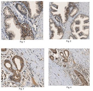
Immunohistochemistry Analysis: A 1:260 dilution from a representative lot detected sFRP-1 in human kideny and testis tissue sections (Courtesy of Jeffrey Rubin, M.D., Ph.D., and Aykut Üren, M.D., Ph.D.).

Neutralizing Analysis: A representative lot, in combination with recombinant Wtn5a and an anti-DKK1 antibody, increased migration and mammosphere formation of EpCAM-positive (EPC+) human mammary epithelial cells (MECs) (Scheel, C., et al. (2011). Cell. 145(6):926 940).

Neutralizing Analysis: A representative lot enhanced the formation of TRACP+ mutinucleated and mononucleated cells from co-cultured murine splenic and primary osteoblastic cells upon PGE2 and 1 ,25 (OH)2D3 stimulatation (Häusler, K.D., et al. (2004). J. Bone Miner. Res. 19(11):1873-1881).

Note: Detection of endogenous sFRP-1 in whole cell lysates or tissue samples is difficult without an enrichment step prior to immunoblotting (e.g. by immunoprecipitation or heparin affinity chromatography).
Research Category
Signaling

Quality

Evaluated by Western Blotting of human sFRP-1 recombinant protein.

Western Blotting Analysis: 1 µg/mL of this antibody detected in 10 ng of mammalian expressed, secreted human sFRP-1.

Target description

~39 kDa observed. Target band size appears larger than the calculated molecular weights of 32.54/35.39 kDa (mature/pro-form) due to glycosylation. Uncharacterized bands may be observed in some lysate(s).

Physical form

Format: Purified
Protein A purified.
Purified rabbit polyclonal antibody in PBS without preservatives.

Storage and Stability

Stable for 1 year at -20°C from date of receipt.
Handling Recommendations: Upon receipt and prior to removing the cap, centrifuge the vial and gently mix the solution. Aliquot into microcentrifuge tubes and store at -20°C. Avoid repeated freeze/thaw cycles, which may damage IgG and affect product performance.

Other Notes

Concentration: Please refer to lot specific datasheet.

Disclaimer

Unless otherwise stated in our catalog or other company documentation accompanying the product(s), our products are intended for research use only and are not to be used for any other purpose, which includes but is not limited to, unauthorized commercial uses, in vitro diagnostic uses, ex vivo or in vivo therapeutic uses or any type of consumption or application to humans or animals.

Not finding the right product?  

Try our Product Selector Tool.

Storage Class Code

12 - Non Combustible Liquids

WGK

WGK 2

Flash Point(F)

Not applicable

Flash Point(C)

Not applicable


Certificates of Analysis (COA)

Search for Certificates of Analysis (COA) by entering the products Lot/Batch Number. Lot and Batch Numbers can be found on a product’s label following the words ‘Lot’ or ‘Batch’.

Already Own This Product?

Find documentation for the products that you have recently purchased in the Document Library.

Visit the Document Library

Mitchell E Fane et al.
Nature, 606(7913), 396-405 (2022-06-02)
Disseminated cancer cells from primary tumours can seed in distal tissues, but may take several years to form overt metastases, a phenomenon that is termed tumour dormancy. Despite its importance in metastasis and residual disease, few studies have been able

Our team of scientists has experience in all areas of research including Life Science, Material Science, Chemical Synthesis, Chromatography, Analytical and many others.

Contact Technical Service