Saltar al contenido
Merck
Todas las fotos(1)

Documentos

MABF937

Sigma-Aldrich

Anti-Notch 3 Antibody, ECD, clone 2G8

clone 2G8, from rat

Sinónimos:

Neurogenic locus notch homolog protein 3, Notch 3, Notch homolog 3, Notch 3 ECD

Iniciar sesiónpara Ver la Fijación de precios por contrato y de la organización


About This Item

UNSPSC Code:
12352203
eCl@ss:
32160702
NACRES:
NA.41

biological source

rat

Quality Level

antibody form

purified immunoglobulin

antibody product type

primary antibodies

clone

2G8, monoclonal

species reactivity

human

technique(s)

immunofluorescence: suitable
immunohistochemistry: suitable (paraffin)
western blot: suitable

isotype

IgG2aκ

NCBI accession no.

UniProt accession no.

shipped in

wet ice

target post-translational modification

unmodified

General description

Neurogenic locus notch homolog protein 3 (UniProt Q9UM47; also known as Notch 3, Notch homolog 3) is encoded by the NOTCH3 (also known as CASIL, CADASIL, IMF2) gene (Gene ID 4854) in human. Notch 3 is a transmembrane receptor required for arterial differentiation and maturation of vascular smooth muscle cells in small arteries. Notch 3 is initially produced with a signal peptide (a.a. 1-39), the removal of which yields the 2282-amino acid Notch 3 (a.a. 40-2321), which undergoes a constitutive proteolytical cleavage, generating the extracellular fragment (Notch3-ECD; a.a. 40-1628) and the transmembrane/cytosolic (Notch3-TMIC; a.a. 1629-2321) fragment that remain associated at the cell surface to form a heterodimeric receptor. The ECD fragment contains all 34 EGF-like domains and the three Lin/Notch (LNR) repeats, while the extracellular portion of TMIC contains 5 ankyrin repeats. Mutations in the NOTCH3 gene have been identified as the underlying cause of cerebral autosomal dominant arteriopathy with subcortical infarcts and leukoencephalopathy (CADASIL). Pathogenic mutations predominantly affect cysteine residues within EGF-like repeats of Notch3-ECD, leading to Notch3-ECD multimerization and accumulation in the tunica media of vessel walls.

Specificity

Clone 2G8 reacts with unprocessed Notch 3 and the extracellular (Notch 3-ECD; a.a. 40-1628) fragment, but not the transmembrane/cytosolic fragment (Notch 3-TMIC, also called Notch 3 extracellular truncation; a.a. 1629-2321) or the Notch 3 intracellular fragment (a.a. 1662-2321).

Immunogen

Epitope: Extracellular EGF-like domains 33-34.
KLH-conjugated linear peptide corresponding to a sequence from the EGF-like domains 33-34 region of human Notch 3 ECD.

Application

Anti-Notch 3 Antibody, ECD, clone 2G8 is an antibody against Notch 3 for use in Immunohistochemistry (Paraffin), Immunofluorescence, Western Blotting.
Immunohistochemistry Analysis: 20 µg/mL of this antibody from a representative lot detected Notch 3 extracellular domain (ECD) deposits in blood vessels of CADASIL patients brain tissue sections (Courtesy of Institute for Stroke and Dementia Research, Clinical center of the Ludwig-Maximilians University Munich, Germany).
Immunohistochemistry Analysis: A representative lot detected the aggregated Notch 3 extracellular domain (ECD) deposits in brain blood vessels of CADASIL patients by immunohistochemistry staining of paraformaldehyde-fixed, paraffin-embedded brain tissue sections (Kast, J., et al. (2014). Acta Neuropathol. Commun. 2:96).
Immunofluorescence Analysis: A representative lot detected the aggregated Notch 3 extracellular domain (ECD) deposits in brain blood vessels of CADASIL patients by fluorescent immunohistochemistry staining of paraformaldehyde-fixed frozen brain tissue sections. The Notch3-EDC immunoreactivity was seen co-localized with that of LTBP-1, but not fibrillin-1 (Kast, J., et al. (2014). Acta Neuropathol. Commun. 2:96).
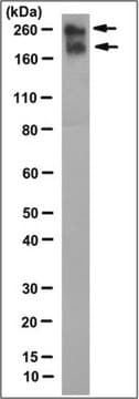
Western Blotting Analysis: A representative lot detected an elevated accumulation of aggregated Notch 3 extracellular domain (ECD) in brain blood vessel extracts from CADASIL patients when comparing to vessel extracts from non-diseased brains (Kast, J., et al. (2014). Acta Neuropathol. Commun. 2:96).
Research Category
Inflammation & Immunology
Research Sub Category
Nervous System

Quality

Identity Confirmation by Isotyping Test.

Isotyping Analysis: The identity of this monoclonal antibody is confirmed by isotyping test to be IgG2aκ.

Target description

243.6 kDa (Pre-form), 239.5 kDa (mature, unprocessed), 167.13 kDa (Notch 3-ECD) calculated. ~250 kDa (Notch 3-ECD) reported (Kast, J., et al. (2014). Acta Neuropathol. Commun. 2:96).

Physical form

Format: Purified
Protein G Purified
Purified rat monoclonal IgG2aκ antibody in buffer containing 0.1 M Tris-Glycine (pH 7.4), 150 mM NaCl with 0.05% sodium azide.

Storage and Stability

Stable for 1 year at 2-8°C from date of receipt.

Other Notes

Concentration: Please refer to lot specific datasheet.

Disclaimer

Unless otherwise stated in our catalog or other company documentation accompanying the product(s), our products are intended for research use only and are not to be used for any other purpose, which includes but is not limited to, unauthorized commercial uses, in vitro diagnostic uses, ex vivo or in vivo therapeutic uses or any type of consumption or application to humans or animals.

Not finding the right product?  

Try our Herramienta de selección de productos.

Storage Class

12 - Non Combustible Liquids

wgk_germany

WGK 1

flash_point_f

Not applicable

flash_point_c

Not applicable


Certificados de análisis (COA)

Busque Certificados de análisis (COA) introduciendo el número de lote del producto. Los números de lote se encuentran en la etiqueta del producto después de las palabras «Lot» o «Batch»

¿Ya tiene este producto?

Encuentre la documentación para los productos que ha comprado recientemente en la Biblioteca de documentos.

Visite la Librería de documentos

Nuestro equipo de científicos tiene experiencia en todas las áreas de investigación: Ciencias de la vida, Ciencia de los materiales, Síntesis química, Cromatografía, Analítica y muchas otras.

Póngase en contacto con el Servicio técnico