Saltar al contenido
Merck
Todas las fotos(2)

Documentos

MAB4423

Sigma-Aldrich

Anti-SOX-2 Antibody, clone 10H9.1

clone 10H9.1, from mouse

Sinónimos:

ANOP3, MCOPS3, MGC2413

Iniciar sesiónpara Ver la Fijación de precios por contrato y de la organización


About This Item

UNSPSC Code:
12352203
eCl@ss:
32160702
NACRES:
NA.41

biological source

mouse

antibody form

purified immunoglobulin

antibody product type

primary antibodies

clone

10H9.1, monoclonal

species reactivity

human, mouse

technique(s)

immunocytochemistry: suitable
western blot: suitable

isotype

IgG2bκ

NCBI accession no.

UniProt accession no.

shipped in

wet ice

target post-translational modification

unmodified

Gene Information

human ... SOX2(6657)
mouse ... Sox2(20674)

General description

SOX 2 is also known as SRY related HMG BOX gene 2. All SOX proteins have a single HMG box and bind linear DNA in a sequence-specific manner, resulting in the bending of DNA through large angles. Bending causes the DNA helix to open for some distance, which may affect binding and interactions of other transcription factors. SOX1, SOX2 and SOX3 show the closest homology to SRY. They share maximum homology within the HMG domain and are expressed mainly in the developing nervous system of the mouse. These genes share significant homology outside the HMG box also and are highly conserved throughout their evolution. Sox-2 plays a key role in embryonic stem cell development, and has been shown to help stem cells maintain a pluripotent potential. It has also been shown to be associated with uncommitted dividing stem cell and precursor cells of the developing CNS.

Immunogen

Recombinant protein corresponding to human SOX-2.

Application

Anti-SOX-2 Antibody, clone 10H9.1 detects level of SOX-2 & has been published & validated for use in WB & IC.
Research Category
Stem Cell Research
Research Sub Category
Pluripotent & Early Differentiation

Neural Stem Cells
Western Blot Analysis: 1.0 µg/mL from a representative lot detected SOX-2 in H9 human stem cell lysate.

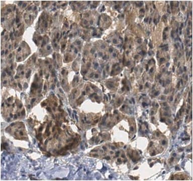
Immunocytochemistry Analysis: 10 µg/mL from a representative lot detected SOX-2 in human RenCX cells.

Quality

Evaluated by Western Blot in mouse embryonic stem cell lysate.

Western Blot Analysis: 0.5 µg/mL of this antibody detected SOX-2 in mouse embryonic stem cell lysate.

Target description

~34 kDa observed

Physical form

Format: Purified
Protein G Purified
Purified mouse monoclonal IgG2bκ in buffer containing 0.1 M Tris-Glycine (pH 7.4), 150 mM NaCl with 0.05% sodium azide.

Storage and Stability

Stable for 1 year at 2-8°C from date of receipt.

Analysis Note

Control
Mouse embryonic stem cells

Other Notes

Concentration: Please refer to the Certificate of Analysis for the lot-specific concentration.

Disclaimer

Unless otherwise stated in our catalog or other company documentation accompanying the product(s), our products are intended for research use only and are not to be used for any other purpose, which includes but is not limited to, unauthorized commercial uses, in vitro diagnostic uses, ex vivo or in vivo therapeutic uses or any type of consumption or application to humans or animals.

¿No encuentra el producto adecuado?  

Pruebe nuestro Herramienta de selección de productos.

Optional

Referencia del producto
Descripción
Precios

Storage Class

12 - Non Combustible Liquids

wgk_germany

WGK 1

flash_point_f

Not applicable

flash_point_c

Not applicable


Certificados de análisis (COA)

Busque Certificados de análisis (COA) introduciendo el número de lote del producto. Los números de lote se encuentran en la etiqueta del producto después de las palabras «Lot» o «Batch»

¿Ya tiene este producto?

Encuentre la documentación para los productos que ha comprado recientemente en la Biblioteca de documentos.

Visite la Librería de documentos

Gurkan Mollaoglu et al.
Immunity, 49(4), 764-779 (2018-10-18)
The major types of non-small-cell lung cancer (NSCLC)-squamous cell carcinoma and adenocarcinoma-have distinct immune microenvironments. We developed a genetic model of squamous NSCLC on the basis of overexpression of the transcription factor Sox2, which specifies lung basal cell fate, and
Aleksandra Beylina et al.
Stem cell research, 53, 102354-102354 (2021-06-06)
Mutations in leucine-rich repeat kinase 2 (LRRK2) are associated with inherited forms of Parkinson's disease (PD), causing disease by a gain of kinase function. Here, we describe a series of isogenic iPSC lines with any of five pathogenic mutations (N1437H
Xiaoyan Zhan et al.
The Journal of experimental medicine, 217(5) (2020-03-18)
Type I interferons (IFNs) are known to mediate antineoplastic effects during tumor progression. Type I IFNs can be produced by multiple cell types in the tumor microenvironment; however, the molecular mechanisms by which tumor cells evade the inhibition of immune
Ana Joana Duarte et al.
Stem cell research, 45, 101794-101794 (2020-05-11)
Fabry Disease (FD) is a multisystemic X-linked disorder that belongs to the group of lysosomal storage disorders (LSDs). Causal mutations on alpha-galactosidase A (α-Gal A) commonly lead to abnormal protein and consequently to FD. Since it is an X-linked disease
Jianghong Man et al.
Cell stem cell, 22(1), 104-118 (2017-12-05)
Tumor hypoxia is associated with poor patient survival and is a characteristic of glioblastoma. Notch signaling is implicated in maintaining glioma stem-like cells (GSCs) within the hypoxic niche, although the molecular mechanisms linking hypoxia to Notch activation have not been

Artículos

Fibroblast growth factors (FGFs) regulate developmental pathways and mesoderm/ectoderm patterning in early embryonic development.

Human iPSC neural differentiation media and protocols used to generate neural stem cells, neurons and glial cell types.

Protocolos

A stem cell culture protocol to generate 3D NSC models of Alzheimer’s disease using ReNcell human neural stem cell lines.

Nuestro equipo de científicos tiene experiencia en todas las áreas de investigación: Ciencias de la vida, Ciencia de los materiales, Síntesis química, Cromatografía, Analítica y muchas otras.

Póngase en contacto con el Servicio técnico