Skip to Content
Merck
All Photos(2)

Documents

06-1433

Sigma-Aldrich

Anti-phospho-FGFR-1 (Tyr653/Tyr654) Antibody

from rabbit, purified by affinity chromatography

Synonym(s):

Basic fibroblast growth factor receptor 1, FGFR-1, bFGF-R-1, Fms-like tyrosine kinase 2, FLT-2, Proto-oncogene c-Fgr, CD331

Sign Into View Organizational & Contract Pricing


About This Item

UNSPSC Code:
12352203
eCl@ss:
32160702
NACRES:
NA.41

biological source

rabbit

Quality Level

antibody form

affinity isolated antibody

antibody product type

primary antibodies

clone

polyclonal

purified by

affinity chromatography

species reactivity

human

species reactivity (predicted by homology)

Xenopus (based on 100% sequence homology), mouse (based on 100% sequence homology), zebrafish (based on 100% sequence homology), chicken (based on 100% sequence homology), bovine (based on 100% sequence homology), rat (based on 100% sequence homology)

packaging

antibody small pack of 25 μg

technique(s)

cell based assay: suitable
western blot: suitable

NCBI accession no.

UniProt accession no.

shipped in

ambient

target post-translational modification

phosphorylation (pTyr653/pTyr654)

Gene Information

human ... FGFR1(2260)

General description

FGFR-1 (fibroblast growth factor receptor 1) is a member of the basic fibroblast growth factor family of proteins and is thought to be important in the induction and propagation of benign prostatic hyperplasia. A shortage of FGFR-1 causes death post-implantation and has been linked to neurofascin and neurite outgrowth. FGFR-1 expression has recently been found to be increased in patients with renal cell carcinoma and FGFR-1 may also have a role to play in early limb formation.

Specificity

This antibody recognizes FGFR-1 phosphorylated at Tyr653/Tyr654.

Immunogen

Epitope: Phosphorylated Tyr653/Tyr654
KLH-conjugated linear peptide corresponding to human FGFR-1 phosphorylated at Tyr653/Tyr654.

Application

Detect phospho-FGFR-1 (Tyr653/Tyr654) using this Anti-phospho-FGFR-1 (Tyr653/Tyr654) Antibody validated for use in WB, Cell Function Assay.
Peptide Inhibition Assay Analysis: 0.2 µg/mL from a representative lot peptide blocked in HEK293T cells transfected with FGFR-1.
Research Category
Inflammation & Immunology
Research Sub Category
Cytokines & Cytokine Receptors

Quality

Evaluated by Western Blot in untreated and lambda phosphatase-treated HEK293T cells transfected with FGFR1.

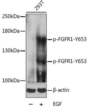
Western Blot Analysis: 0.2 µg/mL of this antibody detected FGFR-1 in 10 µg of untreated and lambda phosphatase-treated HEK293T cells transfected with FGFR1.

Target description

~145 kDa observed.
FGFR-1 has 19 isoforms and eight potential glycosylation sites that increase the expected size to 120 kDa for the immature form and 145 kDa for the mature form of the receptor (Hisaoka, K., et al. (2011). JBC. 286(24):21118-21128).

Physical form

Affinity purified
Purified rabbit polyclonal in buffer containing 0.1 M Tris-Glycine (pH 7.4), 150 mM NaCl with 0.05% sodium azide.

Storage and Stability

Stable for 1 year at 2-8°C from date of receipt.

Analysis Note

Control
Untreated and lambda phosphatase-treated HEK293T cells transfected with FGFR-1

Other Notes

Concentration: Please refer to the Certificate of Analysis for the lot-specific concentration.

Disclaimer

Unless otherwise stated in our catalog or other company documentation accompanying the product(s), our products are intended for research use only and are not to be used for any other purpose, which includes but is not limited to, unauthorized commercial uses, in vitro diagnostic uses, ex vivo or in vivo therapeutic uses or any type of consumption or application to humans or animals.

Not finding the right product?  

Try our Product Selector Tool.


Certificates of Analysis (COA)

Search for Certificates of Analysis (COA) by entering the products Lot/Batch Number. Lot and Batch Numbers can be found on a product’s label following the words ‘Lot’ or ‘Batch’.

Already Own This Product?

Find documentation for the products that you have recently purchased in the Document Library.

Visit the Document Library

Tamer A Ahmed et al.
Cell reports, 26(1), 65-78 (2019-01-04)
Pharmacologic targeting of components of ERK signaling in ERK-dependent tumors is often limited by adaptive resistance, frequently mediated by feedback-activation of RTK signaling and rebound of ERK activity. Here, we show that combinatorial pharmacologic targeting of ERK signaling and the
Jiahui Si et al.
Journal of experimental & clinical cancer research : CR, 38(1), 481-481 (2019-12-06)
Although EGFR tyrosine kinase inhibitors (EGFR-TKIs) are beneficial to lung adenocarcinoma patients with sensitive EGFR mutations, resistance to these inhibitors induces a cancer stem cell (CSC) phenotype. Here, we clarify the function and molecular mechanism of shisa3 as a suppressor
LTBP-2 Has a Single High-Affinity Binding Site for FGF-2 and Blocks FGF-2-Induced Cell Proliferation.
Menz, C; Parsi, MK; Adams, JR; Sideek, MA; Kopecki, Z; Cowin, AJ; Gibson, MA
Testing null
Agata Lampart et al.
Cells, 11(3) (2022-02-16)
Fibroblast growth factor 1 (FGF1) is considered primarily as a ligand for FGF surface receptors (FGFRs) through which it activates a number of cellular responses. In addition to its canonical mode of action, FGF1 can act intracellularly, before secretion or
Mateusz Adam Krzyscik et al.
ACS omega, 2(7), 3792-3805 (2018-07-20)
Antibody-drug conjugates (ADCs) are a new class of anticancer therapeutics that combine the selectivity of targeted treatment, ensured by monoclonal antibodies, with the potency of the cytotoxic agent. Here, we applied an analogous approach, but instead of an antibody, we

Our team of scientists has experience in all areas of research including Life Science, Material Science, Chemical Synthesis, Chromatography, Analytical and many others.

Contact Technical Service