Skip to Content
MilliporeSigma
All Photos(2)

Documents

MAB347

Sigma-Aldrich

Anti-Growth Associated Protein 43 Antibody, clone 9-1E12

clone 9-1E12, Chemicon®, from mouse

Synonym(s):

Neuromodulin, B-50 Protein, Growth Associated Protein 43, neuromodulin

Sign Into View Organizational & Contract Pricing


About This Item

UNSPSC Code:
12352203
eCl@ss:
32160702
NACRES:
NA.41

biological source

mouse

Quality Level

antibody form

purified immunoglobulin

antibody product type

primary antibodies

clone

9-1E12, monoclonal

species reactivity

feline, rat, hamster

species reactivity (predicted by homology)

human

manufacturer/tradename

Chemicon®

technique(s)

immunohistochemistry: suitable
immunoprecipitation (IP): suitable
western blot: suitable

isotype

IgG1

NCBI accession no.

UniProt accession no.

shipped in

wet ice

target post-translational modification

unmodified

Gene Information

human ... GAP43(2596)

General description

GAP-43, a common marker of differentiating neurons, is expressed at elevated levels by developing or regenerating neurons during axon growth. While GAP-43 is an integral membrane protein associated with the cytoplasmic surface of axonal growth cones (and synapses), it is absent from dendritic growth cones. In adult brain, GAP-43 is found in high concentration in presynaptic areas where memory formation is thought to occur, such as frontal cortex, limbic system and hippocampus. Phosphorylation of GAP-43 by PKC (at Ser41 on human, mouse, rat, and bovine GAP-43 or Ser42 on chicken and Xenopus GAP-43) is specifically correlated with certain forms of synaptic plasticity.

Specificity

GAP-43 (also known as growth associated protein-43, B-50, F1 and pp46), regardless of the protein’s phosphorylation state.

Immunogen

GAP-43 purified from rat brain

Application

Anti-Growth Associated Protein 43 Antibody, clone 9-1E12 detects level of Growth Associated Protein 43 & has been published & validated for use in IH, IP & WB.
Immunohistochemistry:
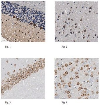
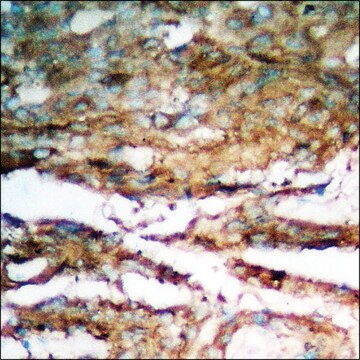
0.1-0.5 µg/mL of a previous lot on 4% paraformaldehyde perfused and fixed rat cerebellum and cerebrum tissue. The addition of 0.01-0.1% Triton X-100 to incubation buffer will increase cellular permeability.

Immunoprecipitation:
A previous lot of this antibody was used in immunoprecipitation.
5-10µg/mL in membrane preparations from 16-18 day embryonic cortical neuronal cultures.

Western Blot Analysis:
MAB347 will detect a single 43-48 kD band in western blots of membrane fractions of growing neurons.

Optimal working dilutions must be determined by the end user.
Research Category
Neuroscience
Research Sub Category
Neuroregenerative Medicine

Quality

Routinely evaluated by Western Blot on rat brain lysates.

Western Blot Analysis:
1:1000 dilution of this lot detected GAP-43/B-50 on 10 μg of rat brain lysates.

Target description

43-48 kDa

Physical form

Format: Purified
Protein A Purified mouse immunoglobulin IgG1 in 20 mM sodium phosphate, 250 mM NaCl, pH. 7.6, with 0.1% sodium azide as a preservative.
Protein A purified

Storage and Stability

Maintain at 2-8°C in undiluted for up to 6 months after date of receipt.

Analysis Note

Control
Rat DRG tissue that has been subjected to a spinal nerve ligation and cultured neurons.

Other Notes

Concentration: Please refer to the Certificate of Analysis for the lot-specific concentration.

Legal Information

CHEMICON is a registered trademark of Merck KGaA, Darmstadt, Germany

Disclaimer

Unless otherwise stated in our catalog or other company documentation accompanying the product(s), our products are intended for research use only and are not to be used for any other purpose, which includes but is not limited to, unauthorized commercial uses, in vitro diagnostic uses, ex vivo or in vivo therapeutic uses or any type of consumption or application to humans or animals.

Not finding the right product?  

Try our Product Selector Tool.

Storage Class

10 - Combustible liquids

wgk_germany

WGK 2

flash_point_f

Not applicable

flash_point_c

Not applicable


Certificates of Analysis (COA)

Search for Certificates of Analysis (COA) by entering the products Lot/Batch Number. Lot and Batch Numbers can be found on a product’s label following the words ‘Lot’ or ‘Batch’.

Already Own This Product?

Find documentation for the products that you have recently purchased in the Document Library.

Visit the Document Library

Faqi Wang et al.
Toxicological sciences : an official journal of the Society of Toxicology, 121(2), 279-291 (2011-03-26)
Perfluorooctane sulfonate (PFOS) and 2,2',4,4'-tetrabromodiphenyl ether (BDE-47) are two persistent environmental contaminants that are toxic to developing nervous systems, particularly via their disruption of thyroid hormone (TH) function. To investigate whether an interaction existed between PFOS and BDE-47 on TH-mediated
Sonia Canterini et al.
The FEBS journal, 280(5), 1320-1329 (2013-01-12)
Proteins of the TSC22 domain (TSC22D) family, including TSC22D1 and TSC22D4, play pivotal roles in cell proliferation, differentiation and apoptosis, interacting with other factors in a still largely unknown manner. This study explores this issue by biochemically characterizing various TSC22D4
Kyu-Hee Han et al.
International journal of molecular medicine, 44(4), 1473-1483 (2019-08-23)
One of the primary theories of the pathogenesis of tinnitus involves maladaptive auditory‑somatosensory plasticity in the dorsal cochlear nucleus (DCN), which is assumed to be due to axonal sprouting. Although a disrupted balance between auditory and somatosensory inputs may occur
Dominik Linz et al.
Circulation. Arrhythmia and electrophysiology, 8(2), 466-474 (2015-02-26)
Atrial fibrillation (AF) leads to structural and neural remodeling in the atrium, which enhances AF complexity and perpetuation. Renal denervation (RDN) can reduce renal and whole-body sympathetic activity. Aim of this study was to determine the effect of sympathetic nervous
Michaela Fredrich et al.
Neural plasticity, 2011, 859359-859359 (2011-12-03)
The matrix metalloproteinases MMP-9 and MMP-2, major modulators of the extracellular matrix (ECM), were changed in amount and distribution in the rat anteroventral cochlear nucleus (AVCN) following its sensory deafferentation by cochlear ablation. To determine what causal relationships exist between

Our team of scientists has experience in all areas of research including Life Science, Material Science, Chemical Synthesis, Chromatography, Analytical and many others.

Contact Technical Service