Skip to Content
MilliporeSigma
All Photos(5)

Documents

06-923

Sigma-Aldrich

Anti-Cdk1/Cdc2 (PSTAIR) Antibody

Upstate®, from rabbit

Synonym(s):

Anti-CDC2

Sign Into View Organizational & Contract Pricing


About This Item

UNSPSC Code:
12352203
eCl@ss:
32160702
NACRES:
NA.41

biological source

rabbit

Quality Level

antibody form

affinity isolated antibody

antibody product type

primary antibodies

clone

polyclonal

species reactivity

human, rat, amphibian, mouse

species reactivity (predicted by homology)

chicken, canine, starfish, monkey, frog, Drosophila, sheep

manufacturer/tradename

Upstate®

technique(s)

immunocytochemistry: suitable
immunoprecipitation (IP): suitable
western blot: suitable

isotype

IgG

NCBI accession no.

UniProt accession no.

shipped in

wet ice

target post-translational modification

unmodified

Gene Information

chicken ... Cdk1(396252)
dog ... Cdk1(100856079)
frog ... Cdk1(394503)
fruit fly ... Cdk1(34411)
human ... CDK1(983)
mouse ... Cdk1(12534)
rat ... Cdk1(54237)

General description

Entry of all eukaryotic cells into mitosis is regulated by activation of cdc2 kinase. Activation of cdc2 is controlled at several steps including cyclin binding and phosphorylation of Thr161. However, the critical regulatory step in activating cdc2 during progression into mitosis appears to be dephosphorylation of Tyr15 and Thr14. Phosphorylation at Tyr15 and inhibition of cdc2 is carried out by Wee1 and Myt1 protein kinases, while Tyr15 dephosphorylation and activation of cdc2 is carried out by the cdc25 phosphatase. Expression of cdc2 plays a critical role in cell transition through the G2/M phase. The activity of cdc2 in dividing cells increases at the onset of M phase coincident with disassembly of the cell nucleus, generation of mitotic spindles and chromosome condensation. Several proteins including Histone H1 and pp60src are substrates for cdc2.

Specificity

Cdk1/cdc2 kinase
Wide species cross-reactivity is expected.

Immunogen

Synthetic peptide corresponding to residues 42-57 of human cdc2 kinase. The immunogen is identical in a wide range of species.

Application

Fluorescent Immunocytochemistry Analysis:
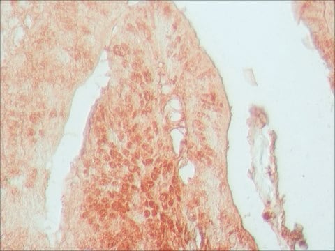
A previous lot of this antibody was used in the confocal Immunofluorescent microscopy of A431 cells. Cells were grown, washed, fixed with formaldahyde, and permeabilized with NP40. The cells were triple stained with with Anti-Cdk1/Cdc2 (Red, Cy3), Phalloidin (Actin, Green, AlexaFluor488), and DAPI (Blue, Nucleus). Positive nuclear staining.

Immunoprecipitation: 4 µg of a previous lot immunoprecipitated cdc2 from 500 µg of A431 RIPA lysate.
Anti-Cdk1/Cdc2 (PSTAIR) Antibody is a high quality Rabbit Polyclonal Antibody for the detection of Cdk1/Cdc2 (PSTAIR) & has been validated in ICC, IP & WB.

Quality

Evaluated by western blot on human A431, mouse 3T3 and/or rat L6 RIPA cell lysates.

Western Blot Analysis: 0.5-2 µg/mL of this antibody detected cdc2 from human A431, mouse 3T3 or rat L6 RIPA cell lysates.

Target description

~34 kDa

Physical form

Format: Purified
Purified rabbit serum in Tris-Glycine (pH 7.4), 150 mM NaCl with 0.05% sodium azide.

Analysis Note

Control
Positive Antigen Control: Catalog #12-301, non-stimulated A431 cell lysate. Add 2.5µL of 2-mercaptoethanol/100µL of lysate and boil for 5 minutes to reduce the preparation. Load 20µg of reduced lysate per lane for mingels.

Legal Information

UPSTATE is a registered trademark of Merck KGaA, Darmstadt, Germany

Not finding the right product?  

Try our Product Selector Tool.

Storage Class

12 - Non Combustible Liquids

wgk_germany

WGK 1

flash_point_f

Not applicable

flash_point_c

Not applicable


Certificates of Analysis (COA)

Search for Certificates of Analysis (COA) by entering the products Lot/Batch Number. Lot and Batch Numbers can be found on a product’s label following the words ‘Lot’ or ‘Batch’.

Already Own This Product?

Find documentation for the products that you have recently purchased in the Document Library.

Visit the Document Library

Cdk1 inhibition induces mutually inhibitory apoptosis and reactivation of Kaposi's sarcoma-associated herpesvirus.
Li, X; Chen, S; Sun, R
Journal of virology null
Tsvetomira Ivanova et al.
Nature communications, 11(1), 2267-2267 (2020-05-10)
To faithfully transmit genetic information, cells must replicate their entire genome before division. This is thought to be ensured by the temporal separation of replication and chromosome segregation. Here we show that in 20-40% of unperturbed yeast cells, DNA synthesis
Platelet-activating factor- and thrombin-induced stimulation of p34cdc2-cyclin histone H1 kinase activity in platelets.
Samiei, M, et al.
The Journal of Biological Chemistry, 266, 14889-14892 (1991)
Tao Fan et al.
Cancer research, 67(16), 7901-7906 (2007-08-19)
A functional genomic approach integrating microarray and proteomic analyses done in our laboratory has identified 14-3-3zeta as a putative oncogene whose activation was common and driven by its genomic amplification in lung adenocarcinomas. 14-3-3zeta is believed to function in cell
Plk1-dependent phosphorylation regulates functions of DNA topoisomerase IIalpha in cell cycle progression.
Li, H; Wang, Y; Liu, X
The Journal of Biological Chemistry null

Our team of scientists has experience in all areas of research including Life Science, Material Science, Chemical Synthesis, Chromatography, Analytical and many others.

Contact Technical Service