
SRE0061
Monoclonal Anti-pan-Cytokeratin
clone PCK-26, Reagents designed and manufactured under cGMP controls suitable for use in an IVD application.
About This Item
Recommended Products


biological source
mouse
conjugate
unconjugated
antibody form
ascites fluid
antibody product type
primary antibodies
clone
PCK-26, monoclonal
contains
15 mM sodium azide
species reactivity
human, chicken, snake, hamster, pig, goat, feline, bovine, carp, rat, rabbit, canine, lizard, sheep, mouse, guinea pig
technique(s)
immunoblotting: suitable
immunohistochemistry (formalin-fixed, paraffin-embedded sections): 1:300
immunohistochemistry (frozen sections): suitable
indirect immunofluorescence: 1:300 using using protease-idgested, formalin-fixed, paraffin-embedded sections of human or animal tissues
isotype
IgG1
shipped in
dry ice
Storage temp.
−20°C
Gene Information
Rattus norvegicus ... Krt1(300250)
bovine ... Krt1(100301161)
dog ... Krt1(444857)
human ... KRT1(3848) , KRT1(3848) , KRT1(3848) , KRT5(3852) , KRT5(3852) , KRT5(3852) , KRT6A(3868) , KRT6A(3868) , KRT6A(3868) , KRT6B(3854) , KRT6B(3854) , KRT6B(3854) , KRT8(3856) , KRT8(3856) , KRT8(3856)
mouse ... KRT1(16678) , KRT1(16678) , KRT1(16678) , Krt1(16678) , Krt5(110308) , Krt5(110308) , Krt5(110308) , Krt6a(16687) , Krt6a(16687) , Krt6a(16687) , Krt6b(16688) , Krt6b(16688) , Krt6b(16688) , Krt8(16691) , Krt8(16691) , Krt8(16691)
rat ... Krt2-5(369017) , Krt2-5(369017) , Krt2-5(369017) , Krt2-8(25626) , Krt2-8(25626) , Krt2-8(25626)
Looking for similar products? Visit Product Comparison Guide
General description
Specificity
Immunogen
Application
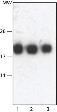
Monoclonal Anti-Cytokeratin, pan may be used for the localization of cytokeratins using various immunochemical assays such as immunoblotting, dot blotting, and immunohistochemstry (immunofluorescence and immunoenzymatic staining).
A minimum antibody titer of 1:300 was determined by indirect immunofluorescent staining of protease digested, formalin-fixed, paraffin-embedded sections of human or animal tissues.
Not finding the right product?
Try our Product Selector Tool.
Storage Class
10 - Combustible liquids
wgk_germany
WGK 3
flash_point_f
Not applicable
flash_point_c
Not applicable
Choose from one of the most recent versions:
Certificates of Analysis (COA)
Don't see the Right Version?
If you require a particular version, you can look up a specific certificate by the Lot or Batch number.
Already Own This Product?
Find documentation for the products that you have recently purchased in the Document Library.
Our team of scientists has experience in all areas of research including Life Science, Material Science, Chemical Synthesis, Chromatography, Analytical and many others.
Contact Technical Service