Skip to Content
MilliporeSigma
All Photos(2)

Key Documents

SAB5500070

Sigma-Aldrich

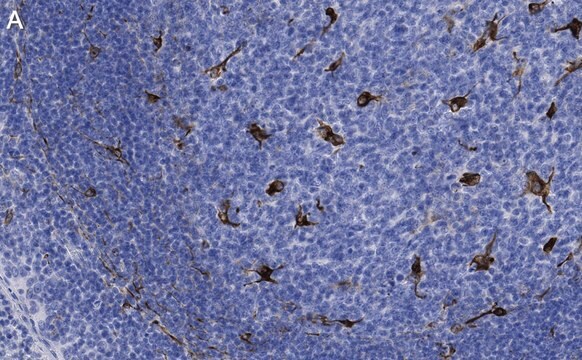
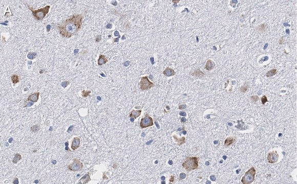
Anti-CD68 antibody, Rabbit monoclonal

clone SP251, recombinant, expressed in proprietary host, affinity isolated antibody

Sign Into View Organizational & Contract Pricing


About This Item

UNSPSC Code:
12352203
NACRES:
NA.41

biological source

rabbit

recombinant

expressed in proprietary host

conjugate

unconjugated

antibody form

affinity isolated antibody

antibody product type

primary antibodies

clone

SP251, monoclonal

species reactivity

human (tested)

technique(s)

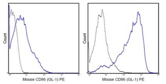
flow cytometry: 1:100
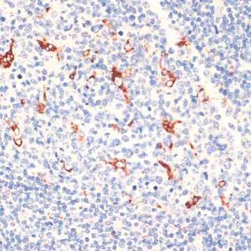
immunohistochemistry: 1:100

isotype

IgG

UniProt accession no.

shipped in

wet ice

Storage temp.

2-8°C

target post-translational modification

unmodified

Gene Information

human ... CD68(968)

General description

CD68 is an intracellular glycoprotein mainly expressed on macrophages and monocytes. It could play a role in phagocytic activities of tissue macrophages, both in intracellular lysosomal metabolism and extracellular cell-cell and cell-pathogen interactions. CD68 is also expressed by interdigitating reticulum cells in tonsil and some histiocytic lymphoma or histiocytosis, acute myeloid leukemia (AML) (M1-M5 types), and granulocytic sarcoma.
CD68 (cluster of differentiation 68) gene is mapped to human chromosome 17p13.1. The gene codes for a glycoprotein, macrosialin. The encoded protein belongs to the family of LAMP (lysosome-associated membrane glycoproteins) proteins. Macrosialin contains a glycosylated mucin-like domain.

Immunogen

Synthetic peptide derived from the internal region of the human CD68 protein.

Application

Monoclonal Anti-CD68 antibody from rabbit has been used for immunohistochemistry.

Biochem/physiol Actions

CD68 (cluster of differentiation 68), a scavenger receptor, significantly participates in the development of Alzheimer′s disease. CD68 serves as a prognostic marker in tumor cells. Deletion of the gene is known to cause reduction in the bone resorption capacity.

Features and Benefits

Evaluate our antibodies with complete peace of mind. If the antibody does not perform in your application, we will issue a full credit or replacement antibody. Learn more.

Physical form

0.1 ml rabbit monoclonal antibody purified by protein A/G in PBS/1% BSA buffer pH 7.6 with less than 0.1% sodium azide.

Disclaimer

Unless otherwise stated in our catalog or other company documentation accompanying the product(s), our products are intended for research use only and are not to be used for any other purpose, which includes but is not limited to, unauthorized commercial uses, in vitro diagnostic uses, ex vivo or in vivo therapeutic uses or any type of consumption or application to humans or animals.

Not finding the right product?  

Try our Product Selector Tool.

Storage Class

12 - Non Combustible Liquids

wgk_germany

WGK 2

flash_point_f

Not applicable

flash_point_c

Not applicable


Choose from one of the most recent versions:

Certificates of Analysis (COA)

Lot/Batch Number

Don't see the Right Version?

If you require a particular version, you can look up a specific certificate by the Lot or Batch number.

Already Own This Product?

Find documentation for the products that you have recently purchased in the Document Library.

Visit the Document Library

Effect of silica oxide nanoparticles on liver of adult male albino rat. Light and electron microscopic study.
Shaimaa M F, et al.
The Egyptian journal of histology., 40(3) (2017)
Multiple Ets factors and interferon regulatory factor-4 modulate CD68 expression in a cell type-specific manner.
O'Reilly D, et al.
The Journal of Biological Chemistry, 278(24), 21909-21919 (2003)
CD68/macrosialin: not just a histochemical marker.
Chistiakov D A, et al.
Laboratory Investigation; a Journal of Technical Methods and Pathology, 97(1), 4-4 (2017)
Jan Van Slambrouck et al.
EBioMedicine, 92, 104608-104608 (2023-05-25)
SARS-CoV-2 is a single-stranded positive-sense RNA virus. Several negative-sense SARS-CoV-2 RNA species, both full-length genomic and subgenomic, are produced transiently during viral replication. Methodologies for rigorously characterising cell tropism and visualising ongoing viral replication at single-cell resolution in histological sections
Elena D Czyrnik et al.
Frontiers in oncology, 13, 1212585-1212585 (2023-09-06)
Cell-cell communication is an important process in healthy tissue but also gains enhanced attention regarding pathological tissue. To date, the tumor microenvironment is gradually brought into focus when studying tumorigenesis. In the prostate gland, stromal and epithelial cells greatly interact

Questions

Reviews

No rating value

Active Filters

Our team of scientists has experience in all areas of research including Life Science, Material Science, Chemical Synthesis, Chromatography, Analytical and many others.

Contact Technical Service