Skip to Content
MilliporeSigma
All Photos(1)

Documents

SAB4200794

Sigma-Aldrich

Anti-Involucrin antibody, Mouse monoclonal

clone SY5, purified from hybridoma cell culture

Synonym(s):

Anti-IVL

Sign Into View Organizational & Contract Pricing


About This Item

UNSPSC Code:
12352203
NACRES:
NA.41

biological source

mouse

Quality Level

antibody form

purified from hybridoma cell culture

antibody product type

primary antibodies

clone

SY5, monoclonal

form

buffered aqueous solution

mol wt

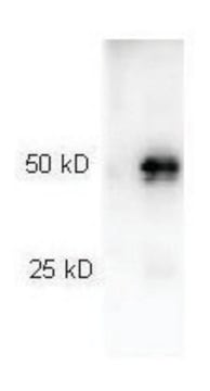
~68 kDa

species reactivity

canine, gorilla, porcine, mouse, human, owl monkey

concentration

~1 mg/mL

technique(s)

immunoblotting: suitable
immunofluorescence: suitable
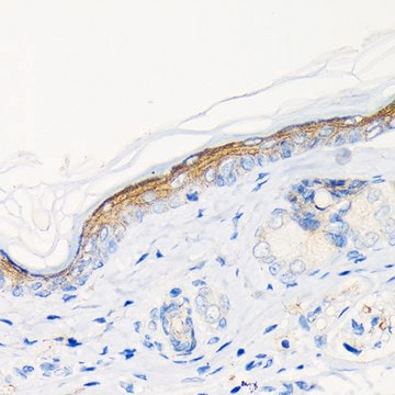
immunohistochemistry: 2.5-5 μg/mL using human skin sections
immunoprecipitation (IP): suitable

isotype

IgG1

UniProt accession no.

shipped in

dry ice

storage temp.

−20°C

target post-translational modification

unmodified

Gene Information

human ... IVL(3713)

General description

Monoclonal Anti-Involucrin (mouse IgG1 isotype) is derived from the SY5 hybridoma, produced by the fusion of mouse myeloma cells and splenocytes from a mouse immunized with purified human involucrin. Involucrin is a soluble cytoplasmic protein. It is expressed in a range of stratified squamous epithelia, including the cornea, which lacks a distinct cornified layer.
The skin serves as a vital barrier that protects the internal environment from external influences. As cells start to migrate out of the basal cell layer of the epidermis towards the surface of the skin, it lose the ability to divide and progress towards a terminal differentiation stage. During this process, keratinocytes go through marked morphological and structural changes. At the same time, the synthesis of important differentiation-dependent structural and catalytic proteins is initiated, which include keratins, filaggrin, transglutaminase, loricrin and involucrin. Involucrin (IVL) is a soluble cytoplasmic protein precursor of the epidermal cornified envelope that becomes crosslinked by transglutaminase during envelope assembly. Involucrin is expressed in a range of stratified squamous epithelia, including the cornea, which lacks a distinct cornified layer. It serves as an early marker of keratinocyte differentiation and a component of the cornified envelope. It is specifically expressed in the suprabasal layers of the epidermis. Involucrin accumulates in the spinous and granular layers as a non-crosslinked precursor and becomes covalently crosslinked to other proteins and lipids to form the cornified envelope scaffolding during the final stages of keratinocyte differentiation. Cell that have lost their ability to divide still continue to express Involucrin.

In pathological conditions, involucrin expression may be altered. In psoriasis and other benign epidermal hyperplasias, involucrin expression appears closer to the basal layer than it′s normal expression. In squamous cell carcinomas and premalignant lesions, involucrin is abnormally increases and found to be reduced in severe dysplasias of the larynx and cervix.

Monoclonal Anti-Involucrin specifically recognizes human and mouse Involucrin. The antibody does not react with mouse epidermis, thus enabling the use of the antibody for study of human xenografts in nude mice. The antibody stains the upper spinous and granular layers in human skin and the cytoplasm of suprabasal terminally differentiated keratinocytes in stratified colonies.

Immunogen

purified human involucrin

Application

Anti-Involucrin antibody, Mouse monoclonal is recommended to use in various immunological techniques, including:
  • Immunoblot
  • Immunofluorescence
  • Immunohistochemistry
  • Immunoprecipitation

Biochem/physiol Actions

Involucrin (IVL) acts as a precursor of the epidermal cornified envelope that becomes crosslinked by transglutaminase during envelope assembly. Involucrin accumulates in the spinous and granular layers as a non-crosslinked precursor and becomes covalently crosslinked to other proteins and lipids to form the cornified envelope scaffolding during the final stages of keratinocyte differentiation. Cells that have lost their ability to divide express involucrin. In pathological conditions, involucrin expression may be altered. In psoriasis and other benign epidermal hyperplasias, involucrin expression appears closer to the basal layer than its normal expression. In squamous cell carcinomas and premalignant lesions, involucrin is abnormally increased and found to be reduced in severe dysplasias of the larynx and cervix.

Physical form

Supplied as a solution in 0.01 M phosphate buffered saline, pH 7.4, containing 15 mM sodium azide.

Other Notes

This product is for R&D use only, not for drug, household, or other uses.

Not finding the right product?  

Try our Product Selector Tool.

Storage Class

12 - Non Combustible Liquids

wgk_germany

WGK 1


Certificates of Analysis (COA)

Search for Certificates of Analysis (COA) by entering the products Lot/Batch Number. Lot and Batch Numbers can be found on a product’s label following the words ‘Lot’ or ‘Batch’.

Already Own This Product?

Find documentation for the products that you have recently purchased in the Document Library.

Visit the Document Library

Mice deficient in involucrin, envoplakin, and periplakin have a defective epidermal barrier
Sevilla LM, et al.
The Journal of Cell Biology, 179, 1599-1599 (2007)
Michael L Samulevich et al.
International journal of molecular sciences, 23(22) (2022-11-27)
Intrinsically disordered proteins (IDPs) move through an ensemble of conformations which allows multitudinous roles within a cell. Keratinocytes, the predominant cell type in mammalian epidermis, have had only a few individual proteins assessed for intrinsic disorder and its possible contribution

Our team of scientists has experience in all areas of research including Life Science, Material Science, Chemical Synthesis, Chromatography, Analytical and many others.

Contact Technical Service