Skip to Content
MilliporeSigma
All Photos(1)

Documents

P5872

Sigma-Aldrich

Anti-Phosphotyrosine antibody, Mouse monoclonal

clone PT-66, purified from hybridoma cell culture

Synonym(s):

Monoclonal Anti-Phosphotyrosine, Phospho-Tyr, Phospho-tyrosine, p-Tyr

Sign Into View Organizational & Contract Pricing


About This Item

UNSPSC Code:
12352203
NACRES:
NA.44

biological source

mouse

Quality Level

conjugate

unconjugated

antibody form

purified immunoglobulin

antibody product type

primary antibodies

clone

PT-66, monoclonal

form

buffered aqueous solution

packaging

antibody small pack of 25 μL

concentration

~2 mg/mL

technique(s)

flow cytometry: suitable
immunocytochemistry: suitable
immunohistochemistry: suitable
immunoprecipitation (IP): suitable
indirect ELISA: 0.5-1.0 μg/mL using phosphotyrosine conjugated to BSA
radioimmunoassay: suitable
western blot: 0.25-0.5 μg/mL using total cell extract of human platelets

isotype

IgG1

shipped in

dry ice

storage temp.

−20°C

target post-translational modification

unmodified

Looking for similar products? Visit Product Comparison Guide

General description

As determined by ELISA and competitive ELISA, the antibody reacts specifically with phosphorylated tyrosine, both as free amino acid or conjugated to carriers such as BSA or KLH. No cross-reactivity is observed with non-phosphorylated tyrosine, phosphothreonine, phosphoserine, AMP or ATP.
Monoclonal Anti-Phosphotyrosine (mouse IgG1 isotype) is derived from the PT-66 hybridoma produced by the fusion of mouse myeloma cells and splenocytes from BALB/c mice immunized with phosphotyrosine-BSA conjugate.

Specificity

Monoclonal Anti-Phosphotyrosine is specific for phosphorylated tyrosine both as the free amino acid or when conjugated to carriers such as BSA or KLH. It does not react with non-phosphorylated tyrosine or other phosphorylated amino acids, including serine

Immunogen

phosphotyrosine conjugated to BSA

Application

Monoclonal Anti-Phosphotyrosine has been used in
  • immunoblotting,
  • immunofluorescence,
  • immunohistochemistry
  • immunocytochemistry
  • flow cytometry
  • immunoprecipitation
  • enzyme linked immunosorbent assay (ELISA)
  • radio immunoassay (RIA)
  • immunoaffinity isolation

Biochem/physiol Actions

Protein phosphorylation is a basic signaling mechanism that modifies protein function in eukaryotic cells. Serine, threonine, and tyrosine are the major phosphorylated amino acids in proteins. Tyrosine phosphorylation is a rare post-translational event in normal tissues, accounting for only 0.03% of phosphorylated amino acids. However, this phosphorylation increases several folds by various activation signals and the process is mediated by protein tyrosine kinases. Protein-tyrosine kinases (PTKs) are enzymes that catalyze the transfer of γ-phosphate of ATP to tyrosine residues of protein substrates. The PTKs are responsible for many biological processes like cell cycle, proliferation, oncogenesis, and development.

Physical form

Solution in 0.01 M phosphate buffered saline, pH 7.4, containing 15 mM sodium azide.

Disclaimer

Unless otherwise stated in our catalog or other company documentation accompanying the product(s), our products are intended for research use only and are not to be used for any other purpose, which includes but is not limited to, unauthorized commercial uses, in vitro diagnostic uses, ex vivo or in vivo therapeutic uses or any type of consumption or application to humans or animals.

Not finding the right product?  

Try our Product Selector Tool.

Storage Class

12 - Non Combustible Liquids

wgk_germany

WGK 2

flash_point_f

Not applicable

flash_point_c

Not applicable


Certificates of Analysis (COA)

Search for Certificates of Analysis (COA) by entering the products Lot/Batch Number. Lot and Batch Numbers can be found on a product’s label following the words ‘Lot’ or ‘Batch’.

Already Own This Product?

Find documentation for the products that you have recently purchased in the Document Library.

Visit the Document Library

Cyclin D1 governs adhesion and motility of macrophages
Neumeister P, et al.
Molecular Biology of the Cell, 14(5), 2005-2015 (2003)
Dongwon Choi et al.
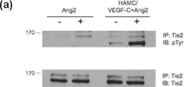
Circulation research, 120(9), 1426-1439 (2017-02-09)
Lymphatic vessels function to drain interstitial fluid from a variety of tissues. Although shear stress generated by fluid flow is known to trigger lymphatic expansion and remodeling, the molecular basis underlying flow-induced lymphatic growth is unknown. We aimed to gain
Phase II study evaluating 2 dosing schedules of oral foretinib (GSK1363089), cMET/VEGFR2 inhibitor, in patients with metastatic gastric cancer
Shah MA, et al.
Testing, 8(3), e54014-e54014 (2013)
alpha1 Integrin cytoplasmic domain is involved in focal adhesion formation via association with intracellular proteins
LOSTER K, et al.
The Biochemical Journal, 356(1), 233-240 (2001)
Autoregulatory mechanisms in protein-tyrosine kinases
Hubbard SR, et al.
The Journal of Biological Chemistry, 273(20), 11987-11990 (1998)

Our team of scientists has experience in all areas of research including Life Science, Material Science, Chemical Synthesis, Chromatography, Analytical and many others.

Contact Technical Service