GBL112358
Grace Bio-Labs CultureWell™ removable chambered coverglass
size 16 wells , Round
Synonym(s):
removable chambered coverglass, removable chambered coverslip
Sign Into View Organizational & Contract Pricing
All Photos(3)
About This Item
UNSPSC Code:
41122600
NACRES:
NB.22
Recommended Products
material
colorless
silicone (on glass)
sterility
sterile
feature
wells 16
packaging
pack of 8 ea
manufacturer/tradename
Grace Bio-Labs 112358
external L × W × depth
80.7 mm × 20.5 mm × 13.7 mm
size
16 wells , Round
well area
0.4 cm2
General description
CS16 - CultureWell™ with Removable Chambered Coverglass - ideal for cell culture and fluorescence imaging applications
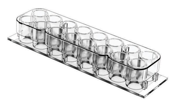
The 2 x 8, 16 chambered coverglass is like a chambered slide with the imaging advantages of a removable coverglass substructure. It is designed with standard 96 well plate volumes and well spacing. The non-cytotoxic silicone well gasket forms a leak-proof seal between the polycarbonate upper structure and the coverglass. CultureWell™ chambered coverglass is shipped sterile and ready to use.
Coverglass removal is facilitated using a very simple device, GBL103259, that reliably separates the parts without the need for excessive force or risk of cover glass breakage. Please order GBL103259 separately.
Silicone removal leaves no residue. Components are manufactured and assembled with special orientation features for ease of handling and convenient identification or specimen location after well structure removal.
Key product dimensions include:
The 2 x 8, 16 chambered coverglass is like a chambered slide with the imaging advantages of a removable coverglass substructure. It is designed with standard 96 well plate volumes and well spacing. The non-cytotoxic silicone well gasket forms a leak-proof seal between the polycarbonate upper structure and the coverglass. CultureWell™ chambered coverglass is shipped sterile and ready to use.
Coverglass removal is facilitated using a very simple device, GBL103259, that reliably separates the parts without the need for excessive force or risk of cover glass breakage. Please order GBL103259 separately.
Silicone removal leaves no residue. Components are manufactured and assembled with special orientation features for ease of handling and convenient identification or specimen location after well structure removal.
- The growth surface is No. 1.5 German borosilicate coverglass
- Inert, non-cytotoxic silicone permits complete edge-to-edge growth of cells
- Well spacing allows use of multi-channel pipettes for ease and speed of handling
- Cell growth characteristics are optimal due to the use of inert silicone materials and manufacturing process
- Quality tested for non-leakage, cell growth and coverglass removal to ensure consistent performance
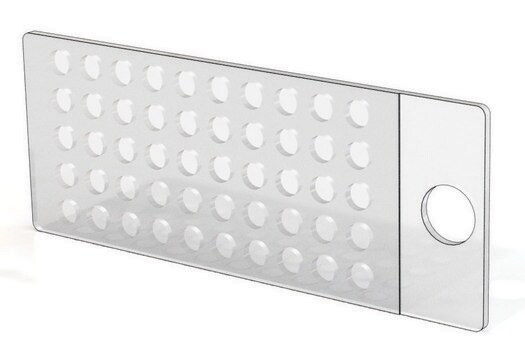
- Silicone well gasket remains attached to coverglass after separation, allowing wells to be used as reagent reservoirs
- Gasket well design is ideally suited for small volume incubations; in situ hybridization and immunostaining
- Black well gasket reduces light scatter, enhancing fluorescence applications. The gasket can easily be removed from the coverglass for mounting
Key product dimensions include:
- Coverslip gasket thickness is 0.5 mm
- Interior diameter of each well is approximately 6 mm
- Volume per gasket well (upper structure removed) is approximately 9 μL
- Volume per well with the upper structure is 250 μL
Application
Cell Culture
Fluorescence applications
In-situ hybridization
Small volume incubation
Immunostaining
Fluorescence applications
In-situ hybridization
Small volume incubation
Immunostaining
Features and Benefits
- The growth surface is No. 1.5
- German borosilicate coverglass
- Inert, non-cytotoxic silicone permits complete edge-to-edge growth of cells
- Well spacing allows use of multi-channel pipettes for ease and speed of handling
- Cell growth characteristics are optimal due to the use of inert silicone materials and manufacturing process
- Quality tested for non-leakage, cell growth and coverglass removal to ensure consistent performance
- Silicone well gasket remains attached to coverglass after separation, allowing wells to be used as reagent reservoirs
- Gasket well design is ideally suited for small volume incubations; in situ hybridization and immunostaining
- Black well gasket reduces light scatter, enhancing fluorescence applications.
- The gasket can easily be removed from the coverglass for mounting
Legal Information
Culturewell is a trademark of Grace Bio-Labs, Inc.
required but not provided
Product No.
Description
Pricing
Certificates of Analysis (COA)
Search for Certificates of Analysis (COA) by entering the products Lot/Batch Number. Lot and Batch Numbers can be found on a product’s label following the words ‘Lot’ or ‘Batch’.
Already Own This Product?
Find documentation for the products that you have recently purchased in the Document Library.
Customers Also Viewed
Yohei Katoh et al.
Molecular biology of the cell, 31(20), 2195-2206 (2020-07-30)
Primary cilia are microtubule-based protrusions from the cell surface that are approximately 0.3 µm in diameter and 3 µm in length. Because size approximates the optical diffraction limit, ciliary structures at the subdiffraction level can be observed only by using
Kyeongbae Min et al.
Methods (San Diego, Calif.), 174, 3-10 (2019-07-22)
Super-resolution microscopy techniques have been widely adopted in biological sciences. Recently, a new super-resolution microscopy technique, called expansion microscopy (ExM) has been developed. In this technique, biomolecules inside specimens are first labeled with fluorophores, followed by in-situ hydrogel synthesis and
Our team of scientists has experience in all areas of research including Life Science, Material Science, Chemical Synthesis, Chromatography, Analytical and many others.
Contact Technical Service


