Skip to Content
MilliporeSigma
All Photos(1)

Documents

F0791

Sigma-Aldrich

Monoclonal Anti-Fibronectin antibody produced in mouse

clone IST-3, ascites fluid

Sign Into View Organizational & Contract Pricing


About This Item

MDL number:
UNSPSC Code:
12352203
NACRES:
NA.41

biological source

mouse

Quality Level

conjugate

unconjugated

antibody form

ascites fluid

antibody product type

primary antibodies

clone

IST-3, monoclonal

mol wt

antigen 240 kDa

contains

15 mM sodium azide

species reactivity

goat, canine, human, chicken, sheep, rabbit, bovine

technique(s)

immunocytochemistry: suitable
immunohistochemistry (frozen sections): suitable
indirect ELISA: suitable
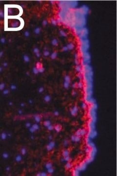
indirect immunofluorescence: 1:50 using cultured human fibroblasts
microarray: suitable
radioimmunoassay: suitable
western blot: suitable

isotype

IgG1

UniProt accession no.

shipped in

dry ice

storage temp.

−20°C

target post-translational modification

unmodified

Gene Information

human ... FN1(2335)

Looking for similar products? Visit Product Comparison Guide

General description

Fibronectin (FN) is an extracellular matrix protein composed of two nearly-identical disulfide-bound polypeptides with typical molecular weights of 220-280 kDa. Cellular fibronectin is structurally and antigenically similar to cold insoluble globulin from plasma and antibodies to either form usually cross-react. Careful analysis of the FN molecule indicates that it contains several functionally and structurally distinct domains which may bind to cell surfaces and to a variety of molecules such as collagen, heparin, gelatin, fibrin and DNA. FN′s play an important role in diverse biological phenomena including cell adhesion, cell migration, hemostasis and thrombosis, wound healing and the ability to induce a more normal phenotype in transformed cells.
Monoclonal Anti-Human Fibronectin (mouse IgG1 iso-type) is derived from the IST-3 hybridoma produced by the fusion of mouse myeloma cells and splenocytes from BALB/c immunized mice. Fibronectin (FN) is a multifunctional extracellular matrix protein comprising two nearly-identical disulfide-bound polypeptides. It is an adhesive glycoprotein. FN belongs to the ligand glycoprotein family and is is expressed in various cell types. Human fibronectin consists of five distinct domains that are referred to as heparin (Hep)-1/ fibrin (Fib)-1, gelatin (Gel), cell surface (Cell), Hep-2 and Fib-2, based on their affinity for Hep, Gel, Cell or Fib. The FN gene is mapped to human chromosome location 2q35.

Specificity

Monoclonal Anti-Human Fibronectin, clone IST-3, recognizes an epitope located within the fourth type-three repeat of human plasma fibronectin.

Immunogen

human plasma fibronectin

Application

Mouse monoclonal clone IST-3 anti-Human Fibronectin antibody has been used in:
  • enzyme linked immuno sorbent assay (ELISA)
  • western blotting
  • dot blot
  • radio immunoassay (RIA)
  • immunocytochemistry
  • immunohistochemistry
  • immunoprecipitation
  • immunofluorescence

Biochem/physiol Actions

Fibronectin (FN) binds to cell surfaces and a variety of molecules, such as collagen, heparin, gelatin, fibrin, and DNA. It plays an important role in diverse biological phenomena including cell adhesion, cell migration, hemostasis, and thrombosis. FN is also involved in wound healing and stimulates a normal phenotype in transformed cells. Upregulation of FN leads to tumors, such as nasopharyngeal carcinoma (NPC), ovarian cancer, and head and neck squamous cell carcinoma.

Physical form

The product is provided as ascites fluid containing 0.1% sodium azide as preservative.

Storage and Stability

For continuous use, store at 2-8 °C for up to one month. For extended storage, the solution may be frozen in working aliquots. Repeated freezing and thawing is not recommended. Storage in "frost-free" freezers not recommended. If slight turbidity occurs upon prolonged storage, clarify the solution by centrifugation before use.

Disclaimer

Unless otherwise stated in our catalog or other company documentation accompanying the product(s), our products are intended for research use only and are not to be used for any other purpose, which includes but is not limited to, unauthorized commercial uses, in vitro diagnostic uses, ex vivo or in vivo therapeutic uses or any type of consumption or application to humans or animals.

Not finding the right product?  

Try our Product Selector Tool.

Storage Class

10 - Combustible liquids

wgk_germany

nwg

flash_point_f

Not applicable

flash_point_c

Not applicable


Certificates of Analysis (COA)

Search for Certificates of Analysis (COA) by entering the products Lot/Batch Number. Lot and Batch Numbers can be found on a product’s label following the words ‘Lot’ or ‘Batch’.

Already Own This Product?

Find documentation for the products that you have recently purchased in the Document Library.

Visit the Document Library

Sahel N Abduljauwad et al.
Scientific reports, 9(1), 5935-5935 (2019-04-13)
Cancer metastasis results from the suppression of adhesion between cancer cells and the extracellular matrix, causing their migration from the primary tumor location and the subsequent formation of tumors in distant organs. This study demonstrates the potential use of nano-sized
Cellular distribution of invadopodia is regulated by nanometer scale surface
protein patterns
Ayaz GB, et al.
European Journal of Cell Biology, 673-684 (2017)
P Castellani et al.
The Journal of cell biology, 103(5), 1671-1677 (1986-11-01)
Fibronectin molecules are dimers composed of subunits whose primary structures may differ. This is due to alternative splicing in at least two regions (ED and IIICS) of the pre-mRNA. Using two monoclonal antibodies specific for two different epitopes of domain
L Zardi et al.
European journal of biochemistry, 146(3), 571-579 (1985-02-01)
Human plasma fibronectin is composed of at least five distinct domains which we refer to as Hep-1/Fib-1, Gel, Cell, Hep-2 and Fib-2 depending on their affinity for heparin (Hep), gelatin (Gel), the cell surface (Cell) or fibrin (Fib). These domains
Xinglong Zheng et al.
The Journal of biological chemistry, 278(32), 30136-30141 (2003-06-07)
ADAMTS13 consists of a reprolysin-type metalloprotease domain followed by a disintegrin domain, a thrombospondin type 1 motif (TSP1), Cys-rich and spacer domains, seven more TSP1 motifs, and two CUB domains. ADAMTS13 limits platelet accumulation in microvascular thrombi by cleaving the

Our team of scientists has experience in all areas of research including Life Science, Material Science, Chemical Synthesis, Chromatography, Analytical and many others.

Contact Technical Service