Skip to Content
MilliporeSigma
All Photos(1)

Documents

C-12295

Sigma-Aldrich

Human Uterine Microvascular Endothelial Cells (HUtMEC)

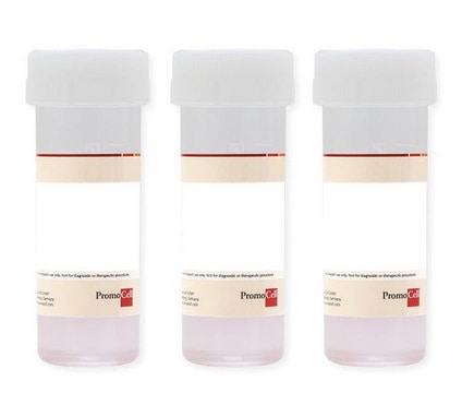
500,000 cryopreserved cells

Sign Into View Organizational & Contract Pricing


About This Item

UNSPSC Code:
41106514
NACRES:
NA.81

biological source

human uterus (myometrium)

packaging

pkg of 500,000 cells

morphology

( endothelial)

technique(s)

cell culture | mammalian: suitable

shipped in

dry ice

storage temp.

−196°C

General description

Lot specific orders are not able to be placed through the web. Contact your local sales rep for more details.

Cell Line Origin

Uterus

Application

Primary Human Uterine Microvascular Endothelial Cells (HUtMEC) are isolated from the myometrium (middle layer of the uterine wall) from a single donor who is not pre-treated with hormones. The cells are provided in a cryopreserved format. The cells are routinely analyzed by immunofluorescent staining: they stain positive for CD31 and von Willebrand factor and negative for smooth muscle alpha-actin. During pregnancy HUtMEC propagate intensely and change their phenotype and expression pattern in response to sex steroids. This includes up-regulation of HUtMEC nitric oxide synthase activity, modulation of adhesion molecule expression, and an increase in endothelial cell survival and angiogenic activity. HUtMEC are involved in medical conditions like uterine fibroma or adenomysis.

Quality

Rigid quality control tests are performed for each lot of Microvascular Endothelial Cells. They are tested for cell morphology and cell type specific markers, e.g. von Willebrand Factor (vWF), CD31 and Podoplanin using flow cytometric analyses. Growth performance is tested through multiple passages up to 15 population doublings (PD) without antibiotics or antimycotics. In addition, all cells have been tested forthe absence of HIV-1, HIV-2, HBV, HCV, HTLV-1, HTLV-2 and microbial contaminants (fungi, bacteria, and mycoplasma).

Warning

Although tested negative for HIV-1, HIV-2, HBV, HCV, HTLV-1 and HTLV-2, the cells – like all products of human origin – should be handled as potentially infectious. No test procedure can completely guarantee the absence of infectious agents.

Subculture Routine

Click here for more information.

Other Notes

Recommended Plating Density: 10000 - 20000 cells per cm2Passage After Thawing: P2Tested Markers: von Willebrand Factor (vWF) positive, CD31 positive, Dil-Ac-LDL uptake positiveGuaranteed population doublings: >15

Recommended products

Recommended Primary Cell Culture Media:Link

Disclaimer

RESEARCH USE ONLY. This product is regulated in France when intended to be used for scientific purposes, including for import and export activities (Article L 1211-1 paragraph 2 of the Public Health Code). The purchaser (i.e. enduser) is required to obtain an import authorization from the France Ministry of Research referred in the Article L1245-5-1 II. of Public Health Code. By ordering this product, you are confirming that you have obtained the proper import authorization.

Storage Class

12 - Non Combustible Liquids

wgk_germany

WGK 1

flash_point_f

Not applicable

flash_point_c

Not applicable


Certificates of Analysis (COA)

Search for Certificates of Analysis (COA) by entering the products Lot/Batch Number. Lot and Batch Numbers can be found on a product’s label following the words ‘Lot’ or ‘Batch’.

Already Own This Product?

Find documentation for the products that you have recently purchased in the Document Library.

Visit the Document Library

Protocols

Cell culture protocol: the endothelial cell transwell migration and invasion assay used to study angiogenesis and cancer cell metastasis. Explore over 350 PromoCell products.

Related Content

Cell culture protocol: the endothelial tube formation assay to study angiogenesis using HUVECs and other endothelial cell types. Explore over 350 PromoCell products.

Our team of scientists has experience in all areas of research including Life Science, Material Science, Chemical Synthesis, Chromatography, Analytical and many others.

Contact Technical Service