Moesin (membrane-organizing extension spike protein) is the protein encoded by the MSN gene. The MSN encoded protein moesin is a member of the ERM (ezrin, moesin, radixin) family of proteins that regulate linkage between the plasma membrane and the actin cytoskeleton. Thus, the MSN encoded protein is a peripheral membrane protein that is keenly involved in the signaling and structural connections in cytoskeletal plasma membrane structures like microvilli. Moesin in its active, phosphorylated state strongly binds actin and helps anchor it to the plasma membrane. The MSN encoded Moesin is localized to filopodia and other membranous extensions of various cell types and thus plays a role in cell-cell recognition signaling, cell movement, and cell transport. In its inactive, non-phosphorylated state, Moesin binds itself in a head-tail fashion and is incapable of binding actin or other cytoskeletal proteins. The kinase ROCK2 is central to the opening up of moesin and allowing its interaction with actin and the plasma membrane. EMD-Millipore’s Anti-MSN, clone 2C12 has been tested in western blots against purified recombinant protein as well as multiple human cancer cell lysates. The clone has also been tested successfully in paraffin embedded immunohistochemistry on human colon tissue and by flow cytometry on human Jurkat cells and ELISA using purified recombinant antigen.
Immunogen
Purified protein fragment of human MSN expressed in E. coli.
Application
Detect Moesin using this mouse monoclonal antibody, Anti-MSN Antibody, clone 2C12 validated for use in western blotting, IHC & Flow Cytometry.
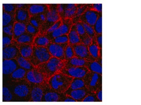
Immunohistochemistry Analysis: A 1:100-1,000 dilution from a representative lot detected MSN in human tonsil and colon tissues.
Flow Cytometry Analysis: A 1:200-400 dilution from a representative lot detected MSN in Jurkat cells.
Optimal working dilutions must be determined by end user.
Quality
Evaluated by Western Blotting in Jurkat cell lysate.
Western Blotting Analysis: A 1:500 dilution of this antibody detected MSN in 10 µg of Jurkat cell lysate.
Target description
~68 kDa observed. Uncharacterized bands may appear in some lysate(s).
Search for Certificates of Analysis (COA) by entering the products Lot/Batch Number. Lot and Batch Numbers can be found on a product’s label following the words ‘Lot’ or ‘Batch’.
Already Own This Product?
Find documentation for the products that you have recently purchased in the Document Library.
Our team of scientists has experience in all areas of research including Life Science, Material Science, Chemical Synthesis, Chromatography, Analytical and many others.