Skip to Content
MilliporeSigma
All Photos(3)

Documents

MABT326

Sigma-Aldrich

Anti-CEACAM3/5 Antibody, clone 308/3-3

clone 308/3-3, from mouse

Synonym(s):

Carcinoembryonic antigen-related cell adhesion molecule 3, Carcinoembryonic antigen CGM1, CD66d, Carcinoembryonic antigen-related cell adhesion molecule 5, Carcinoembryonic antigen, CEA, Meconium antigen 100, CD66e

Sign Into View Organizational & Contract Pricing


About This Item

UNSPSC Code:
12352203
eCl@ss:
32160702
NACRES:
NA.43

biological source

mouse

Quality Level

antibody form

purified antibody

antibody product type

primary antibodies

clone

308/3-3, monoclonal

species reactivity

human

should not react with

rat, mouse

technique(s)

ELISA: suitable
flow cytometry: suitable
immunoprecipitation (IP): suitable
western blot: suitable

isotype

IgG1κ

NCBI accession no.

UniProt accession no.

target post-translational modification

unmodified

Gene Information

human ... CEACAM5(1048)

Related Categories

General description

Carcinoembryonic antigen-related cell adhesion molecule 3 (UniProt P40198; also known as Carcinoembryonic antigen CGM1, CD66d) is encoded by the CEACAM3 (also known as CD66D, CGM1) gene (Gene ID 1084) in human. The carcinoembryonic antigen- (CEA-) related cell adhesion molecules (CEACAMs) constitute a 12-member subgroup of the CEA family of immunoglobulin-related proteins first described in 1965 (PMID 4953873). CEACAMs are reported to participate in diverse physiological processes, including cell adhesion, differentiation, proliferation, and survival, as well as in carcinogenesis and bacterial pathogenesis. CEACAM molecules from neighbouring cells can interact via their respective extracellular N-terminal IgV-like domain and mediate cell-to-cell adhesion through trans-oligomerization. CEACAM molecules within the same cell can also undergo transmembrane domain-mediated cis-oligomerization, an event important for sustaining downstream cellular signaling. Athough CEACAM1 is shown to utilize its cytoplasmic domain for transducing cellular signaling, not all CEACAM members are expressed with a significant cytoplasmic domain, and others (CEACAM5/6/7/8) are GPI-anchored without even a transmembrane domain. Human CEACAM3 and CEACAM4 are two CEACAMs whose extracellular region is composed of only one V-type Ig-like extracellular domain (a.a. 35-142 of CEACAM3). Human CEACAM5 (UniProt P06731), on the other hand, is a GPI-anchored CEACAM with the most extracellular Ig domains, 7 in all.

Specificity

Clone 308/3-3 reacts with human CEACAM3 and CEACAM5, but not human CEACAM1/4/6/7/8, rat CEACAM1, or mouse CEACAM1 & CEACAM2.

Immunogen

Epitope: Extracellular domain.
Human IgG1 Fc-tagged recombinant human CEACAM3 extracellular domain.

Application

Anti-CEACAM Antibody, clone 308/3-3 is an antibody against CEACAM3/5 for use in Western Blotting, ELISA, Flow Cytometry, Immunoprecipitation.
ELISA Analysis: 10 μg/mL from a representative lot detected CHO cell-expressed full-length glycosylated human CEACAM3 and CEACAM5, but not CEACAM1 (CEACAM1-4L), CEACAM4, CEACAM6, CEACAM7, or CEACAM8 (Courtesy of Dr. Bernhard B. Singer, University of Duisburg-Essen, Germany).
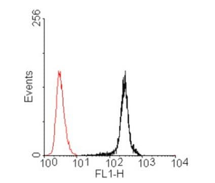
Flow Cytometry Analysis: 10 μg/mL from a representative lot immunostained CHO cell transfectants expressing human CEACAM3 and CEACAM5, but not CHO cells expressing human CEACAM1 (CEACAM1-4L), CEACAM4, CEACAM6, CEACAM7, or CEACAM8 (Courtesy of Dr. Bernhard B. Singer, University of Duisburg-Essen, Germany).
Immunoprecipitation Analysis: 5 μg from a representative lot immunoprecipitated exogenously expressed full-length glycosylated human CEACAM5 from 500 μg of lysate (in 600 μL) from transfected HeLa cells (Courtesy of Dr. Bernhard B. Singer, University of Duisburg-Essen, Germany).
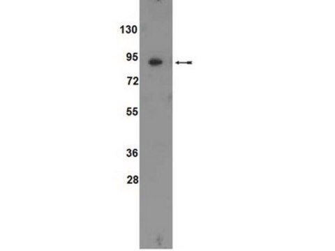
Western Blotting Analysis: 10 μg/mL from a representative lot detected exogenously expressed full-length glycosylated human CEACAM5 in 50 μg of lysate from transfected HeLa cells (Courtesy of Dr. Bernhard B. Singer, University of Duisburg-Essen, Germany).
Research Category
Cell Structure
Research Sub Category
Adhesion (CAMs)

Quality

Evaluated by Western Blotting Analysis in lysates of CEACAM3 & CEACAM5 transfected CHO cells.

Western Blotting Analysis: 2.0 μg/mL of this antibody detected exogenously expressed full-length glycosylated human CEACAM3 and CEACAM5 in 10 μg of lysates from transfected CHO cells.

Target description

~30/180 kDa (CEACAM3/CEACAM5) observed. 27.09/19.40/22.75 kDa (human CEACAM3 isoform CGM1A/CGM1B/CGM1C) and 76.80/76.72 kDa (human CEACAM5 isoform 1/2) calculated. Target bands appear smeary and larger than the calculated molecular weights due to glycosylation. Uncharacterized band(s) may appear in some lysates.

Physical form

Format: Purified
Protein G Purified
Purified mouse monoclonal IgG1κ in buffer containing 0.1 M Tris-Glycine (pH 7.4), 150 mM NaCl with 0.05% sodium azide.

Storage and Stability

Stable for 1 year at 2-8°C from date of receipt.

Other Notes

Concentration: Please refer to lot specific datasheet.

Disclaimer

Unless otherwise stated in our catalog or other company documentation accompanying the product(s), our products are intended for research use only and are not to be used for any other purpose, which includes but is not limited to, unauthorized commercial uses, in vitro diagnostic uses, ex vivo or in vivo therapeutic uses or any type of consumption or application to humans or animals.

Not finding the right product?  

Try our Product Selector Tool.

Storage Class

12 - Non Combustible Liquids

wgk_germany

WGK 1

flash_point_f

Not applicable

flash_point_c

Not applicable


Certificates of Analysis (COA)

Search for Certificates of Analysis (COA) by entering the products Lot/Batch Number. Lot and Batch Numbers can be found on a product’s label following the words ‘Lot’ or ‘Batch’.

Already Own This Product?

Find documentation for the products that you have recently purchased in the Document Library.

Visit the Document Library

Our team of scientists has experience in all areas of research including Life Science, Material Science, Chemical Synthesis, Chromatography, Analytical and many others.

Contact Technical Service