Skip to Content
MilliporeSigma
All Photos(1)

Documents

MAB5212

Sigma-Aldrich

Anti-Neurocan Antibody, clone 650.24

clone 650.24, Chemicon®, from mouse

Synonym(s):

245 kDa Early Postnatal Core Glycoprotein

Sign Into View Organizational & Contract Pricing


About This Item

UNSPSC Code:
12352203
eCl@ss:
32160702
NACRES:
NA.41

biological source

mouse

Quality Level

antibody form

purified immunoglobulin

antibody product type

primary antibodies

clone

650.24, monoclonal

species reactivity

rat

manufacturer/tradename

Chemicon®

technique(s)

immunocytochemistry: suitable
immunohistochemistry: suitable
immunoprecipitation (IP): suitable
western blot: suitable

isotype

IgG1

NCBI accession no.

UniProt accession no.

shipped in

wet ice

target post-translational modification

unmodified

Gene Information

human ... NCAN(1463)

General description

Neurocan is the major soluble chondroitin sulfate proteoglycan in the brain. It is thought to play a functional role in axonal growth and guidance and in the establishment of specific neural pathways during embryonic brain development. Neurocan expression in the brain is developmentally regulated. Early on the major form of neurocan consists of a 245 kD core protein with approximately two chondroitin sulfate glycosoaminoglycan chains of 22 kD each. Later neurocan comprises a 180 kD core protein. Both forms of neurocan contain only chondroitin 4-sulfate glycosoaminoglycan chains. By virtue of their high expression at sites of neurnal damage and trauma, chondroitin sulfate proteoglycans, including neurocan, are thought to inhibit successful nerve regeneration.

Specificity

Neurocan. The specificity of clone 650.24 was checked by western blots of brain lysates and purified neurocan; the antibody reacted with the identical bands that clones 1D1 and 1F6 (other neurocan monoclonals) reacted; furthermore, the staining pattern of brain sections with these antibodies were all identical. Finally 650.24 also recognized recombinant neurocan expressed in transfected 293 cells by western blot.

Immunogen

Embryonic rat brain proteoglycans

Application

Research Category
Neuroscience
Research Sub Category
Neuroregenerative Medicine

Growth Cones & Axon Guidance
This Anti-Neurocan Antibody, clone 650.24 is validated for use in IP, WB, IC, IH for the detection of Neurocan.
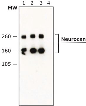
Western blot: 1-2 μg/mL. Reacts with polypeptides of 260 and 160 kDa on western blots of embryonic rat brain tissue extracts. The 160 kDa species is typically only seen aftyer chondroitinase treatment.Treatment is at a concentration of chondroitinase of 10U/mL in Tris-HCL pH 8.0. Make tissue or cell extract in 20-50mM Tris pH 7.6-8.0 with 0.15M NaCl in the presence of protease inhibitors. Add 1 microliter of enzyme to 30 microliters of extract and incubate 30 minutes at 37C. Then add SDS sample buffer, heat or boil sample as normal for SDS reducing samples.

Immunocytochemistry: 1:5

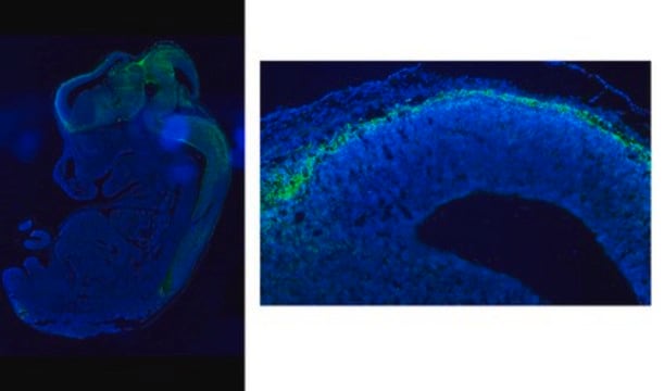
Immunohistochemistry on 4% paraformaldehyde fixed tissue: 1:1,000

Immunoprecipitation: 1-2 μg/mL

Optimal working dilutions must be determined by the end user.

Physical form

Format: Purified
Purified immunoglobulin. Liquid in 0.02M Phosphate buffer, 0.25M NaCl with 0.1% sodium azide.

Storage and Stability

Maintain at 2-8°C in undiluted aliquots for up to 6 months.

Analysis Note

Control
POSITIVE CONTROL:

early post-natal rat brain. Not expressed in kidney, lung, liver, or muscle.

Other Notes

Concentration: Please refer to the Certificate of Analysis for the lot-specific concentration.

Legal Information

CHEMICON is a registered trademark of Merck KGaA, Darmstadt, Germany

Disclaimer

Unless otherwise stated in our catalog or other company documentation accompanying the product(s), our products are intended for research use only and are not to be used for any other purpose, which includes but is not limited to, unauthorized commercial uses, in vitro diagnostic uses, ex vivo or in vivo therapeutic uses or any type of consumption or application to humans or animals.

Not finding the right product?  

Try our Product Selector Tool.

Storage Class

10 - Combustible liquids

wgk_germany

WGK 2

flash_point_f

Not applicable

flash_point_c

Not applicable


Certificates of Analysis (COA)

Search for Certificates of Analysis (COA) by entering the products Lot/Batch Number. Lot and Batch Numbers can be found on a product’s label following the words ‘Lot’ or ‘Batch’.

Already Own This Product?

Find documentation for the products that you have recently purchased in the Document Library.

Visit the Document Library

Marc R Del Bigio et al.
Cerebrospinal fluid research, 5, 12-12 (2008-07-12)
The cerebral cortex may be compressed in hydrocephalus and some experiments suggest that movement of extracellular substances through the cortex is impaired. We hypothesized that the extracellular compartment is reduced in size and that the composition of the extracellular compartment
Alterations in chondroitin sulfate proteoglycan expression occur both at and far from the site of spinal contusion injury.
Andrews, EM; Richards, RJ; Yin, FQ; Viapiano, MS; Jakeman, LB
Experimental neurology null
Wu-Fu Chen et al.
CNS neuroscience & therapeutics, 21(9), 698-707 (2015-07-21)
To date, no reliable methods have proven effective for treating spinal cord injury (SCI). Even systemic administration of methylprednisolone (MP) remains controversial. We previously reported that intrathecal (i.t.) administration of granulocyte colony-stimulating factor (G-CSF) improves outcome after experimental spinal cord
James M Massey et al.
Experimental neurology, 209(2), 426-445 (2007-06-02)
Increased chondroitin sulfate proteoglycan (CSPG) expression in the vicinity of a spinal cord injury (SCI) is a primary participant in axonal regeneration failure. However, the presence of similar increases of CSPG expression in denervated synaptic targets well away from the
Frauke Seehusen et al.
PloS one, 11(7), e0159752-e0159752 (2016-07-22)
In demyelinating diseases, changes in the quality and quantity of the extracellular matrix (ECM) may contribute to demyelination and failure of myelin repair and axonal sprouting, especially in chronic lesions. To characterize changes in the ECM in canine distemper demyelinating

Our team of scientists has experience in all areas of research including Life Science, Material Science, Chemical Synthesis, Chromatography, Analytical and many others.

Contact Technical Service