Skip to Content
MilliporeSigma
All Photos(2)

Documents

MAB13170

Sigma-Aldrich

Anti-Cav1.2 calcium channel Antibody, clone L57/46

clone L57/46, from mouse

Synonym(s):

calcium channel, voltage-dependent, L type, alpha 1C subunit1, voltage-gated calcium channel alpha subunit Cav1.2, calcium channel, L type, alpha 1 polypeptide, isoform 1, cardic muscle, calcium channel, cardic dihydropyridine-sensitive, alpha-1 subunit,

Sign Into View Organizational & Contract Pricing


About This Item

UNSPSC Code:
12352203
eCl@ss:
32160702
NACRES:
NA.41

biological source

mouse

Quality Level

antibody form

purified immunoglobulin

antibody product type

primary antibodies

clone

L57/46, monoclonal

species reactivity

mouse, human

species reactivity (predicted by homology)

rabbit (based on 100% sequence homology), rat (based on 100% sequence homology)

technique(s)

immunohistochemistry: suitable
western blot: suitable

isotype

IgG2bκ

NCBI accession no.

UniProt accession no.

shipped in

wet ice

target post-translational modification

unmodified

Gene Information

General description

This gene encodes an alpha-1 subunit of a voltage-dependent calcium channel. Calcium channels mediate the influx of calcium ions into the cell upon membrane polarization. The alpha-1 subunit consists of 24 transmembrane segments and forms the pore through which ions pass into the cell. The calcium channel consists of a complex of alpha-1, alpha-2/delta, beta, and gamma subunits in a 1:1:1:1 ratio. There are multiple isoforms of each of these proteins, either encoded by different genes or the result of alternative splicing of transcripts. The protein encoded by this gene binds to and is inhibited by dihydropyridine. Many alternate transcriptional splice variants of this gene have been observed but have not been thoroughly characterized.

Specificity

This antibody recognizes Cav1.2 calcium channel at the cytoplasmic domain.

Immunogen

Epitope: Cytoplasmic domain
Recombinant protein corresponding to rabbit Cav1.2 calcium channel at the cytoplasmic domain proximal to the C-terminus.

Application

Immunohistochemistry Analysis: 1:300 dilution of this antibody has been shown to detect Cav1.2 calium channel in human kidney tissue.
Research Category
Neuroscience
Research Sub Category
Ion Channels & Transporters
This Anti-Cav1.2 calcium channel Antibody, clone L57/46 is validated for use in WB, IH for the detection of Cav1.2 calcium channel.

Quality

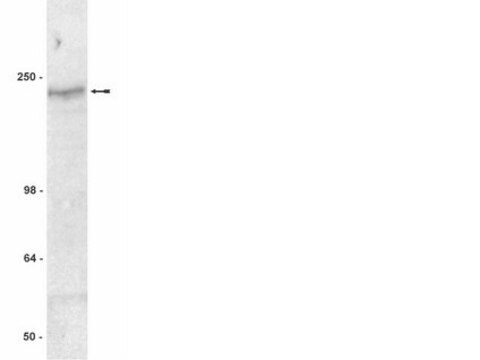
Evaluated by Western Blot in mouse brain tissue lysate.

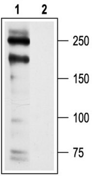
Western Blot Analysis: 2 µg/mL of this antibody detected Cav1.2 calcium channel on 10 µg of mouse brain tissue lysate.

Target description

Observed at ~ 240 kDa

Physical form

Format: Purified
Protein G Purified
Purified mouse monoclonal IgG2bκ in buffer containing 0.1 M Tris-Glycine (pH 7.4, 150 mM NaCl) with 0.05% sodium azide.

Storage and Stability

Stable for 1 year at 2-8°C from date of receipt.

Analysis Note

Control
Mouse brain tissue lysate

Other Notes

Concentration: Please refer to the Certificate of Analysis for the lot-specific concentration.

Disclaimer

Unless otherwise stated in our catalog or other company documentation accompanying the product(s), our products are intended for research use only and are not to be used for any other purpose, which includes but is not limited to, unauthorized commercial uses, in vitro diagnostic uses, ex vivo or in vivo therapeutic uses or any type of consumption or application to humans or animals.

Not finding the right product?  

Try our Product Selector Tool.

Storage Class

12 - Non Combustible Liquids

wgk_germany

WGK 1

flash_point_f

Not applicable

flash_point_c

Not applicable


Certificates of Analysis (COA)

Search for Certificates of Analysis (COA) by entering the products Lot/Batch Number. Lot and Batch Numbers can be found on a product’s label following the words ‘Lot’ or ‘Batch’.

Already Own This Product?

Find documentation for the products that you have recently purchased in the Document Library.

Visit the Document Library

Jia Li et al.
FEBS letters, 596(24), 3145-3158 (2022-08-04)
Cardiomyopathies are ascribed to a variety of etiologies, present with diverse clinical phenotypes, and lack disease-modifying treatments. Mounting evidence implicates dysregulated activin receptor signaling in heart disease and highlights inhibition of this pathway as a potential therapeutic target. Here, we
Christine Y Ivashchenko et al.
American journal of physiology. Heart and circulatory physiology, 305(6), H913-H922 (2013-07-09)
Human-induced pluripotent stem cell-derived cardiomyocytes (hiPS-CMs) have been recently derived and are used for basic research, cardiotoxicity assessment, and phenotypic screening. However, the hiPS-CM phenotype is dependent on their derivation, age, and culture conditions, and there is disagreement as to
Dianaly T Au et al.
Arteriosclerosis, thrombosis, and vascular biology, 38(11), 2651-2664 (2018-10-26)
Objective- Mutations affecting contractile-related proteins in the ECM (extracellular matrix), microfibrils, or vascular smooth muscle cells can predispose the aorta to aneurysms. We reported previously that the LRP1 (low-density lipoprotein receptor-related protein 1) maintains vessel wall integrity, and smLRP1-/- mice
Elizabeth J Akin et al.
The Journal of general physiology, 155(11) (2023-09-13)
Pulmonary arterial (PA) smooth muscle cells (PASMC) generate vascular tone in response to agonists coupled to Gq-protein receptor signaling. Such agonists stimulate oscillating calcium waves, the frequency of which drives the strength of contraction. These Ca2+ events are modulated by
Christopher J Garland et al.
Science signaling, 10(486) (2017-07-06)
Vascular smooth muscle contraction is suppressed by feedback dilation mediated by the endothelium. In skeletal muscle arterioles, this feedback can be activated by Ca2+ signals passing from smooth muscle through gap junctions to endothelial cells, which protrude through holes in

Our team of scientists has experience in all areas of research including Life Science, Material Science, Chemical Synthesis, Chromatography, Analytical and many others.

Contact Technical Service