
MAB1277
Anti-Nucleoli Antibody, clone 125-10
clone 125-10, Chemicon®, from mouse
Sign Into View Organizational & Contract Pricing
All Photos(1)
About This Item
UNSPSC Code:
12352203
eCl@ss:
32160702
NACRES:
NA.41
Recommended Products


biological source
mouse
antibody form
purified from hybridoma cell culture
antibody product type
primary antibodies
clone
125-10, monoclonal
species reactivity
human
manufacturer/tradename
Chemicon®
technique(s)
immunofluorescence: suitable
immunohistochemistry: suitable
isotype
IgG1
shipped in
wet ice
target post-translational modification
unmodified
Specificity
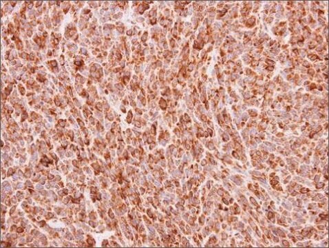
Gives diffuse nucleolar staining pattern on all human cell types.
Application
Detect Nucleoli with Anti-Nucleoli Antibody, clone 125-10 (Mouse Monoclonal Antibody), that has been shown to work in IHC & IF.
Indirect immunofluorescence: 1:20-1:30. Use 30-100 μl per well/slide. Optimal working dilutions must be determined by the end user.
Subcellular Particle
Suggested Protocol
IMMUNOFLUORESCENCE AND ANTIBODY SCREENING PROCEDURE
Hybridoma supernatants are examined by indirect immunofluorescence on cell preparations of human lymphnoid cells. In order to examine many samples in a short period of time, washed cells (wash 2 times in wash buffer at 4°C) at a concentration of 5 x 10⁶ cells/mL in PBS are pipetted dropwise on PTFE coated printed microscope slides containing ten 5 mm wells/slide. After the cells are allowed to settle to the surface of the glass (10-15 minutes only), the overlying fluid is quickly removed by aspiration and the cells are dried to the slide by a gentle stream of warm air. The slides are then immediately fixed in 2% formaldehyde, ultra-pure, in PBS for 15 minutes at room temperature. After fixation, the slides are rinsed in PBS and placed in acetone at -20°C for 3 minutes to make the cells permeable. After a final rinse in PBS to remove the acetone, the slides are stored in PBS at 4°C indefinitely in covered Coplan jars.
In addition to the lymphnoid cultures, normal human epithelial cells can be screened by indirect immunofluorescence microscopy for positive reactions with the hybridoma supernatants. Since the human epithelial cells grow as monolayer cultures, they are plated directly onto the printed microscope slides after trypsinization and allowed to attach and grow overnight at 37°C in complete medium. The following day, the slides are briefly rinsed in PBS to remove the medium and the cells are fixed as described above. In general, the slides are not allowed to air dry either during or after the fixation procedure in order to maintain the cellular integrity and antigenicity of intracellular molecules.
For photographic analysis, viable cell preparations obtained from Ficoll®-hypaque gradient separations are cytocentrifuged directly onto slides at 1250 rpm for 10 minutes. This procedure flattens the lymphnoid cells and greatly improves the visibility of intranuclear and cytoplasmic antigens. Slides prepared in this manner are fixed in the same way directly after cytocentrifugation.
In order to screen the hybridoma supernatants by indirect immunofluorescence, 30-100 μL of each supernatant (optimize for each individual assay) are pipetted on a well of the printed microscope slides using a different tip for each supernatant. After 60 minutes of incubation at 37°C in a humidified chamber, the slides are rinsed 3 times with PBS at room temperature, and again incubated for 30 minutes at 37°C with 20 μL of a 1:20 dilution of fluorescein-conjugated goat anti-mouse IgG antibody (Millipore AP124F). The slides are then prepared rinsed 3 times with PBS, counterstained with Evans Blue for 5 minutes at room temperature using a freshly prepared solution containing 50 μL of a 1% stock solution of Evans Blue in 80 mL of PBS, rinsed a final time in PBS, and coverslipped using a 1:1 solution of glycerol: PBS. The slides are then examined by epifluorescence microscopy. Since many of the monoclonal antibodies produced a rapidly diminishing fluorescent reaction, exposure times optimally are less than five seconds.
Important Note: During shipment, small volumes of antibody will occasionally become entrapped in the seal of the product vial. For antibodies with volumes of 200 μl or less, we recommend gently tapping the vial on a hard surface or briefly centrifuging the vial in a tabletop centrifuge to dislodge any liquid in the container′s cap.
Physical form
Format: Purified
Hybridoma supernatant, concentrated by ammonium sulfate. Buffer: PBS with 0.1% sodium azide.
Legal Information
CHEMICON is a registered trademark of Merck KGaA, Darmstadt, Germany
Ficoll is a registered trademark of Cytiva
Not finding the right product?
Try our Product Selector Tool.
Storage Class
10 - Combustible liquids
wgk_germany
WGK 2
flash_point_f
Not applicable
flash_point_c
Not applicable
Certificates of Analysis (COA)
Search for Certificates of Analysis (COA) by entering the products Lot/Batch Number. Lot and Batch Numbers can be found on a product’s label following the words ‘Lot’ or ‘Batch’.
Already Own This Product?
Find documentation for the products that you have recently purchased in the Document Library.
A novel PHF6 mutation results in enhanced exon skipping and mild Borjeson-Forssman-Lehmann syndrome.
Vallee, D; Chevrier, E; Graham, GE; Lazzaro, MA; Lavigne, PA; Hunter, AG; Picketts, DJ
Journal of medical Genetics null
Active Filters
Our team of scientists has experience in all areas of research including Life Science, Material Science, Chemical Synthesis, Chromatography, Analytical and many others.
Contact Technical Service