Skip to Content
MilliporeSigma
All Photos(3)

Key Documents

ABN1660

Sigma-Aldrich

Anti-S-Opsin

from rabbit

Synonym(s):

Short-wave-sensitive opsin 1, Blue cone photoreceptor pigment, Blue-sensitive opsin, BOP, Short wavelength-sensitive cone opsin

Sign Into View Organizational & Contract Pricing


About This Item

UNSPSC Code:
12352203
eCl@ss:
32160702

biological source

rabbit

antibody form

unpurified

antibody product type

primary antibodies

clone

polyclonal

species reactivity

mouse

species reactivity (predicted by homology)

rat (based on 100% sequence homology)

technique(s)

electron microscopy: suitable
immunofluorescence: suitable
immunohistochemistry: suitable (paraffin)
western blot: suitable

isotype

IgG

NCBI accession no.

UniProt accession no.

shipped in

ambient

target post-translational modification

unmodified

Gene Information

mouse ... Opn1Sw(12057)

General description

Short-wave-sensitive opsin 1 (UniProt: P51491; also known as S opsin, Blue cone photoreceptor pigment, Blue-sensitive opsin, BOP, Short wavelength-sensitive cone opsin) is encoded by the Opn1sw (also known as Bcp) gene (Gene ID: 12057) in murine species. Opsins are G-protein coupled protein with photoreceptor activity. Opsins contain seven transmembrane alpha-helical domains connected by three extra-cellular and three cytoplasmic loops. They are light-sensitive proteins found in photoreceptor cells of the retina. Based on sequence homology, the opsins are classified into six subfamilies: the vertebrate opsin/encephalopsin subfamily, the Go opsin subfamily, the Gs opsin subfamily, the invertebrate Gq opsin subfamily, the photoisomerase subfamily, and the neuropsin subfamily. They share less than 20 percent identity between subfamilies. Opsins provide the necessary environment for the absorption of light at a particular wavelength. Therefore, by providing a different opsin to the retinal, organisms can sense light of different wavelengths. S-opsin is a multi-pass membrane protein that is expressed in cone photoreceptor cells. They are light-absorbing molecules that mediate vision and contain an apoprotein, opsin, which is covalently linked to cis-retinal. It can increase spectral sensitivity in dim light.

Specificity

This polyclonal antibody specifically detects short-wave-sensitive opsin 1 in murine species. Murine and human S-opsin display 88% sequence homology. It targets an epitope within 17 amino acids from the C-terminal region.

Immunogen

KLH-conjugated linear peptide corresponding to 17 amino acids from the C-terminal end of murine short-wave-sensitive opsin 1.

Application

Anti-S-Opsin, Cat. No. ABN1660, is a highly specific rabbit polyclonal antibody that targets Short-wave-sensitive opsin 1 and has been tested in Electron Microscopy, Immunofluorescence, Immunohistochemistry (Paraffin), and Western Blotting.
Electron Microscopy Analysis: A representative lot detected S-Opsin in P30 retinas from COP-T/WT and MOP-T/WT mice (Chakraborty, D., et. al. (2009). Hum Mol Genet. 18(5):797-808).

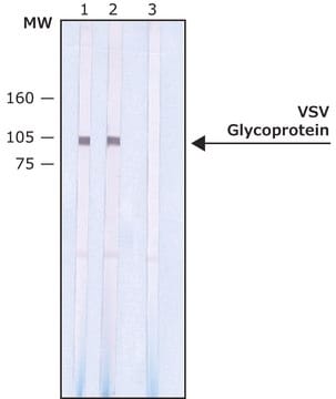
Western Blotting Analysis: A 1:1,000 dilution from a representative lot detected S-Opsin in mouse retinal extract (Courtesy of Dr. Muna Naash at University of Houston, Houston, TX).

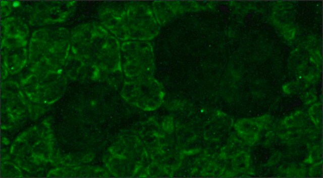
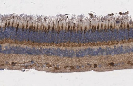
Immunofluorescence Analysis: A representative lot detected S-Opsin in paraffin-embedded retinal and outer segment disc sections from WT and COP-T/WT mice (Chakraborty, D., et. al. (2009). Hum Mol Genet. 18(5):797-808).

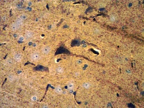
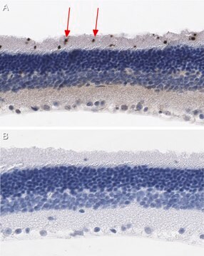
Immunohistochemistry Analysis: A 1:1,000 dilution from a representative lot detected S-Opsin in mouse retina tissue.

Quality

Evaluated by Western Blotting in NRL-/-KO Ms retina lysate.

Western Blotting Analysis: A 1:1,000 dilution of this antibody detected S-Opsin in 5 µg of NRL-/-KO mouse retina lysate.

Target description

~ 37 kDa observed; 38.92 kDa calculated. Uncharacterized bands may be observed in some lysate(s).

Physical form

Format: Unpurified

Other Notes

Concentration: Please refer to lot specific datasheet.

Not finding the right product?  

Try our Product Selector Tool.

Storage Class

12 - Non Combustible Liquids

wgk_germany

WGK 1

flash_point_f

Not applicable

flash_point_c

Not applicable


Certificates of Analysis (COA)

Search for Certificates of Analysis (COA) by entering the products Lot/Batch Number. Lot and Batch Numbers can be found on a product’s label following the words ‘Lot’ or ‘Batch’.

Already Own This Product?

Find documentation for the products that you have recently purchased in the Document Library.

Visit the Document Library

Chun-Lei Deng et al.
Cell death and differentiation, 28(3), 1041-1061 (2020-10-22)
Photoreceptor apoptosis is recognized as one key pathogenesis of retinal degeneration, the counteraction of which represents a promising approach to safeguard visual function. Recently, mesenchymal stem cell transplantation (MSCT) has demonstrated immense potential to treat ocular disorders, in which extracellular
Yogita Kanan et al.
Free radical biology & medicine, 198, 118-122 (2023-02-04)
Retinitis pigmentosa (RP) is caused by many different mutations that promote the degeneration of rod photoreceptors and have no direct effect on cones. After the majority of rods have died cone photoreceptors begin to slowly degenerate. Oxidative damage contributes to
Yuntong Sun et al.
Journal of nanobiotechnology, 21(1), 449-449 (2023-11-25)
Photoreceptor apoptosis is an important pathogenesis of retinal degeneration and a primary cause of vision loss with limited treatment methods. Mesenchymal stem/stromal cells-derived small extracellular vesicles (MSC-sEVs) have shown therapeutic value in various ocular disorders. Recent studies have revealed that
Ashish Bhandari et al.
Frontiers in cellular neuroscience, 13, 426-426 (2019-10-15)
Axonopathy is a hallmark of many neurodegenerative diseases including glaucoma, where elevated intraocular pressure (ocular hypertension, OHT) stresses retinal ganglion cell (RGC) axons as they exit the eye and form the optic nerve. OHT causes early changes in the optic
Xiang Ren et al.
Diabetes research and clinical practice, 185, 109788-109788 (2022-02-20)
Autophagy and exosome secretion in photoreceptor and RPE cells play an important role during diabetic retinopathy (DR). Thioredoxin (Trx) upregulation delays diabetes-induced photoreceptor cell degeneration, which the effect of autophagy and exosome secretion on it is unclear. Therefore, we investigated

Questions

Reviews

No rating value

Active Filters

Our team of scientists has experience in all areas of research including Life Science, Material Science, Chemical Synthesis, Chromatography, Analytical and many others.

Contact Technical Service