Skip to Content
MilliporeSigma
All Photos(1)

Documents

05-723

Sigma-Aldrich

Anti-RhoE/Rnd3 Antibody, clone 4

ascites fluid, clone 4, Upstate®

Synonym(s):

MemB protein, Rho family GTPase 3, ras homolog gene family, member E, small GTP binding protein Rho8

Sign Into View Organizational & Contract Pricing


About This Item

UNSPSC Code:
12352203
eCl@ss:
32160702
NACRES:
NA.41

biological source

mouse

Quality Level

antibody form

ascites fluid

antibody product type

primary antibodies

clone

4, monoclonal

species reactivity

mouse, human

manufacturer/tradename

Upstate®

technique(s)

immunocytochemistry: suitable
western blot: suitable

isotype

IgG

NCBI accession no.

UniProt accession no.

shipped in

dry ice

target post-translational modification

unmodified

Gene Information

human ... RND3(390)

General description

RhoE, also known as Rnd3, (Rho family GTPase 3) is a small G-protein that is a member of the Rnd family of Rho GTPases, which is a part of the Ras superfamily. The activity of Rnd1, Rnd2, and Rnd3/RhoE are not activated by the traditional conformational change of the GTP binding like other Rho family members. As the name dictates, Rnd proteins were associated with making a cell “round”. Like other small G-proteins, the do bind GTP, but unlike other G-proteins, they do not hydrolyze GTP to GDP. The actions of RhoE are associated with the actin cytoskeletal organization in response to external stimuli. RhoE acts antagonistically to RhoA in inducing loss of stress fiber formation, in part by binding to the RhoA downstream kinase Rho-associated kinase (ROCK1). This prevents ROCK1 activation and inhibits downstream signaling. RhoE does go through the post-translation modification of farnylation which enables it to localize and bind to various internal membranes. RhoE is also known to effect cycle progression.

Specificity

This antibody recognizes RhoE/Rnd3, Mr 29 kDa. Weakly cross-reacts with Rnd1, Mr 27 kDa.

Immunogen

Full length fusion protein corresponding to residues 1-244 of mouse RhoE/Rnd3. Clone 4.

Application

Anti-RhoE/Rnd3 Antibody, clone 4 is an antibody against RhoE/Rnd3 for use in IC & WB.
Immunocytochemistry:
A 1:50 dilution of this antibody has been reported by an independent laboratory to show positive immunostaining for RhoE/Rnd3 in 3T3/A31 cells fixed with paraformaldehyde and permeabilized by Triton X-100 (Riento, K., 2003).
Research Category
Signaling
Research Sub Category
Cytoskeletal Signaling

Quality

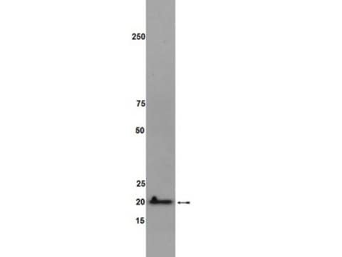
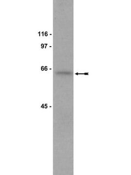
Routinely evaluated by western blot on RIPA lysates from 3T3/A31 cells.

Western Blot Analysis:
A 1:500-1:2000 dilution of this lot detected RhoE/Rnd3 in RIPA lysates from 3T3/A31 cells.

Target description

RhoE/Rnd3 (~29 kDa); Rnd1 (~27 kDa).

Physical form

Mouse monoclonal ascites IgG in buffer containing 0.05% sodium azide before the addition of glycerol to 30%. Liquid at -20ºC.
Unpurified

Storage and Stability

Stable for 1 year at -20°C from date of receipt. For maximum recovery of product, centrifuge the vial prior to removing the cap.

Analysis Note

Control
Positive Antigen Control: Catalog #12-305, 3T3/A31 lysate. Add 2.5 μL of 2-mercapto-ethanol/100 μL of lysate and boil for 5 minutes to reduce the preparation. Load 20 μg of reduced lysate per lane for minigels.

Other Notes

Concentration: Please refer to the Certificate of Analysis for the lot-specific concentration.

Legal Information

UPSTATE is a registered trademark of Merck KGaA, Darmstadt, Germany

Disclaimer

Unless otherwise stated in our catalog or other company documentation accompanying the product(s), our products are intended for research use only and are not to be used for any other purpose, which includes but is not limited to, unauthorized commercial uses, in vitro diagnostic uses, ex vivo or in vivo therapeutic uses or any type of consumption or application to humans or animals.

Not finding the right product?  

Try our Product Selector Tool.

Storage Class

10 - Combustible liquids

wgk_germany

WGK 1


Certificates of Analysis (COA)

Search for Certificates of Analysis (COA) by entering the products Lot/Batch Number. Lot and Batch Numbers can be found on a product’s label following the words ‘Lot’ or ‘Batch’.

Already Own This Product?

Find documentation for the products that you have recently purchased in the Document Library.

Visit the Document Library

Josep Escrivá-Fernández et al.
Journal of biological engineering, 17(1), 34-34 (2023-05-05)
Multiple myeloma (MM) is the second most common hematologic neoplasm which is characterized by proliferation and infiltration of plasmatic cells in the bone marrow. Currently, MM is considered incurable due to resistance to treatment. The CRISPR/Cas9 system has emerged as
Cristina Cueto-Ureña et al.
Frontiers in cell and developmental biology, 10, 834561-834561 (2022-07-15)
Autophagy is a highly conserved process that mediates the targeting and degradation of intracellular components to lysosomes, contributing to the maintenance of cellular homeostasis and to obtaining energy, which ensures viability under stress conditions. Therefore, autophagy defects are common to
A switch in RND3-RHOA signaling is critical for melanoma cell invasion following mutant-BRAF inhibition.
Klein, RM; Higgins, PJ
Molecular Cancer null
Dujuan Cao et al.
Thoracic cancer, 12(7), 1048-1057 (2021-02-11)
Chaperone-mediated autophagy (CMA) is a lysosomal degradation pathway of selective soluble proteins. Lysosomal membrane associated protein 2a (LAMP2a) is the lysosomal membrane receptor of CMA and influences CMA activity. Although it has been suggested that higher expression of LAMP2a is
R Matthew Klein et al.
Cancer research, 69(6), 2224-2233 (2009-02-27)
The depth of cell invasion into the dermis is a clinical determinant for poor prognosis in cutaneous melanoma. The signaling events that promote the switch from a noninvasive to invasive tumor phenotype remain obscure. Activating mutations in the serine/threonine kinase

Our team of scientists has experience in all areas of research including Life Science, Material Science, Chemical Synthesis, Chromatography, Analytical and many others.

Contact Technical Service