Skip to Content
Merck
All Photos(3)

Key Documents

G4405

Sigma-Aldrich

Anti-Guanylyl Cyclase β1 (ER-19) antibody produced in rabbit

enhanced validation

affinity isolated antibody, buffered aqueous solution

Synonym(s):

Anti-GC-S-beta-1, Anti-GC-SB3, Anti-GUC1B3, Anti-GUCB3, Anti-GUCSB3, Anti-GUCY1B3

Sign Into View Organizational & Contract Pricing


About This Item

MDL number:
UNSPSC Code:
12352203
NACRES:
NA.41

biological source

rabbit

Quality Level

conjugate

unconjugated

antibody form

affinity isolated antibody

antibody product type

primary antibodies

clone

polyclonal

form

buffered aqueous solution

mol wt

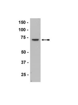
antigen 70 kDa

species reactivity

rat, mouse, human, bovine

enhanced validation

independent
Learn more about Antibody Enhanced Validation

technique(s)

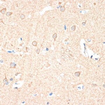
immunohistochemistry (formalin-fixed, paraffin-embedded sections): 1:400 using trypsin-digested human, bovine and mouse heart tissue
immunoprecipitation (IP): 2-3 μg using 60-120 μg of a cytosolic fraction of rat brain
western blot: 1:2,000 using cytosolic fraction of rat brain

shipped in

dry ice

storage temp.

−20°C

target post-translational modification

unmodified

Gene Information

General description

Soluble guanylyl cyclase (sGC) is an obligate hemoprotein enzyme consisting of α and β subunits of ~80 kDa and ~70 kDa, respectively.

Immunogen

synthetic peptide corresponding to amino acid residues 189-207, with N-terminal added lysine, of rat soluble guanylate cyclase 1, conjugated to KLH with glutaraldehyde. The sequence differs in human, bovine and mouse by 1 amino acid.

Application

Anti-Guanylyl Cyclase β1 (ER-19) antibody produced in rabbit is suitable for immunoblotting at a working dilution of 1:2000 using a cytosolic fraction of rat brain, immunoprecipitation at a working antibody amount of 2-3μg using 60-120μg of a cytosolic fraction of rat brain and for immunohistochemistry at 1:400 using trypsin-digested human, bovine and mouse heart tissue.
It has been used as a primary antibody for:
  • localization of β1 subunits of sGC (soluble guanylate cyclase) in the guinea pig gastrointestinal tract
  • detection of expression of sGC in the vasculature of rat skeletal muscle
  • localization of the functional subunit of NO receptors, sGCβ1 in guinea pig caecum
It has also been used in immunoblot assay to determine whether there were changes in uterine vascular smooth muscle (UVSM) sGC expression during the ovine reproductive cycle.

Biochem/physiol Actions

Guanylyl cyclase (GC) catalyzes the conversion of guanosine-5′–triphosphate (GTP) to cyclic guanosine-3′,5-monophosphate (cGMP) and pyrophosphate. This reaction requires Mg2+ or Mn2+. Both the units are required for catalytic activity. The N-terminal domains of the subunits are essential for the stimulation of the enzyme by NO. Dimerization is mediated by the central portion of GC. The C-terminus domain of both subunits forms the catalytic domain.
The enzyme (GC) is a major physiological receptor for nitric oxide (NO), an important intra- and intercellular membrane-permeant signaling molecule. Gaseous NO binds to Fe2+ in the prosthetic heme group of the enzyme. NO binding is followed by disruption of the β1 subunit histidine105 bond to iron and activation of the enzyme. GC forms a complex with NO and cGMP and regulates smooth muscle relaxation, inflammation, platelet adhesion and aggregation, pulmonary physiology and neuronal function. It is an important target for NO-releasing and non-NO-releasing activator drugs in human cardiovascular therapy.

Physical form

Solution in 0.01 M phosphate buffered saline, pH 7.4, containing 1% bovine serum albumin and 15 mM sodium azide.

Disclaimer

Unless otherwise stated in our catalog or other company documentation accompanying the product(s), our products are intended for research use only and are not to be used for any other purpose, which includes but is not limited to, unauthorized commercial uses, in vitro diagnostic uses, ex vivo or in vivo therapeutic uses or any type of consumption or application to humans or animals.

Not finding the right product?  

Try our Product Selector Tool.

Storage Class Code

10 - Combustible liquids

WGK

WGK 1

Flash Point(F)

Not applicable

Flash Point(C)

Not applicable


Certificates of Analysis (COA)

Search for Certificates of Analysis (COA) by entering the products Lot/Batch Number. Lot and Batch Numbers can be found on a product’s label following the words ‘Lot’ or ‘Batch’.

Already Own This Product?

Find documentation for the products that you have recently purchased in the Document Library.

Visit the Document Library

Oxygen Binding and Redox Properties of the Heme in Soluble Guanylate Cyclase IMPLICATIONS FOR THE MECHANISM OF LIGAND DISCRIMINATION
Makino R, et al.
The Journal of Biological Chemistry, 286(18), 15678-15687 (2011)
Allyson G Hindle et al.
American journal of physiology. Regulatory, integrative and comparative physiology, 316(6), R704-R715 (2019-03-21)
Nitric oxide (NO) is a potent vasodilator, which improves perfusion and oxygen delivery during tissue hypoxia in terrestrial animals. The vertebrate dive response involves vasoconstriction in select tissues, which persists despite profound hypoxia. Using tissues collected from Weddell seals at
Staffan Hildebrand et al.
Communications biology, 5(1), 197-197 (2022-03-05)
The nitric oxide-cGMP (NO-cGMP) pathway is of outstanding importance for vascular homeostasis and has multiple beneficial effects in vascular disease. Neointimal hyperplasia after vascular injury is caused by increased proliferation and migration of vascular smooth muscle cells (VSMCs). However, the
Charles R Rosenfeld et al.
American journal of physiology. Heart and circulatory physiology, 296(6), H1878-H1887 (2009-05-28)
Regulation of uteroplacental blood flow (UPBF) during pregnancy remains unclear. Large conductance, Ca(2+)-activated K(+) channels (BK(Ca)), consisting of alpha- and regulatory beta-subunits, are expressed in uterine vascular smooth muscle (UVSM) and contribute to the maintenance of UPBF in the last
Holger Zagst et al.
Analytical and bioanalytical chemistry, 414(4), 1699-1712 (2021-12-07)
Two-dimensional separations provide a simple way to increase the resolution and peak capacity of complex protein separations. The feasibility of a recently developed instrumental approach for two-dimensional separations of proteins was evaluated. The approach is based on the general principle

Our team of scientists has experience in all areas of research including Life Science, Material Science, Chemical Synthesis, Chromatography, Analytical and many others.

Contact Technical Service