Skip to Content
Merck
All Photos(1)

Key Documents

C1399

Sigma-Aldrich

Monoclonal Anti-Cytokeratin Peptide 18 antibody produced in mouse

clone KS-B17.2, ascites fluid

Synonym(s):

Anti-CK18

Sign Into View Organizational & Contract Pricing


About This Item

MDL number:
UNSPSC Code:
12352203
NACRES:
NA.41

biological source

mouse

Quality Level

conjugate

unconjugated

antibody form

ascites fluid

antibody product type

primary antibodies

clone

KS-B17.2, monoclonal

contains

15 mM sodium azide

species reactivity

human

technique(s)

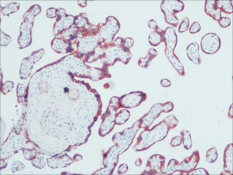
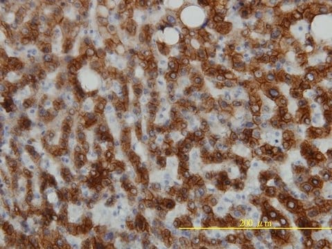
immunohistochemistry (formalin-fixed, paraffin-embedded sections): suitable
indirect immunofluorescence: 1:100 using formalin-fixed, paraffin-embedded sections of human tissue
microarray: suitable

isotype

IgG1

UniProt accession no.

shipped in

dry ice

storage temp.

−20°C

target post-translational modification

unmodified

Gene Information

human ... KRT18(3875)

General description

Monoclonal anti-Cytokeratin Peptide 18 (mouse IgG1 isotype) is derived from the hybridoma produced by the fusion of mouse myeloma cells and splenocytes from an immunized mouse.

Specificity

The antibody reacts with a wide variety of simple epithelia (gastrointestinal, respiratory, urinary, liver, and glandular) but does not react with most stratified squamous epithelium (esophagus, epidermis) or non-epithelial cells.

Immunogen

keratin from the bovine mammary gland epithelial cell line BMGE.

Application

Monoclonal Anti-Cytokeratin Peptide 18 antibody produced in mouse has been used in:
  • immunofluorescence
  • immunoperoxidase staining
  • immunocytochemistry
  • microarray

Biochem/physiol Actions

Cytokeratins are proteins of keratin-containing intermediate filaments that provide mechanical support and other additional functions in epithelial cells. It is found in the intracytoplasmic cytoskeleton of epithelial tissue. Epithelial tissue expresses cytokeratin subunits in a specific and stable pattern. Cytokeratins along with vimentin are involved in cell proliferation, migration and differentiation of preodontoblasts and preameloblasts. The intermediate-sized filaments are abundant in human endothelial cells and are mostly of vimentin type.

Disclaimer

Unless otherwise stated in our catalog or other company documentation accompanying the product(s), our products are intended for research use only and are not to be used for any other purpose, which includes but is not limited to, unauthorized commercial uses, in vitro diagnostic uses, ex vivo or in vivo therapeutic uses or any type of consumption or application to humans or animals.

Not finding the right product?  

Try our Product Selector Tool.

Storage Class Code

10 - Combustible liquids

WGK

nwg

Flash Point(F)

Not applicable

Flash Point(C)

Not applicable


Certificates of Analysis (COA)

Search for Certificates of Analysis (COA) by entering the products Lot/Batch Number. Lot and Batch Numbers can be found on a product’s label following the words ‘Lot’ or ‘Batch’.

Already Own This Product?

Find documentation for the products that you have recently purchased in the Document Library.

Visit the Document Library

Trehalose maintains vitality of mouse epididymal epithelial cells and mediates gene transfer.
Qu B, et al.
PLoS ONE, 9(3), e92483-e92483 (2014)
S K Mohapatra et al.
Animal : an international journal of animal bioscience, 9(4), 643-649 (2014-12-11)
Insulin-like growth factor binding protein-5 (IGFBP-5) is a key molecule in mammary gland development, which facilitates the removal of mammary epithelial cells (MECs) by apoptosis that takes place during remodeling of the mammary gland during involution. IGFBP-5 binds with IGFs
The identification and localization of two intermediate filament proteins in the tunic of Styela plicata (Tunicata, Styelidae).
Di Bella MA, et al.
Tissue & cell, 41(6), 381-389 (2009)
P H Chen et al.
Journal of virology, 67(6), 3507-3514 (1993-06-01)
Immunofluorescence studies revealed that adenovirus induces a reorganization of the cytokeratin system in lytically infected HeLa cells. At 24 h postinfection, the cytokeratin network began to disassemble into prominent spheroid globules. By 36 h postinfection, host cell lysis occurred, accompanied
R Levy et al.
Differentiation; research in biological diversity, 39(3), 185-196 (1988-12-01)
The aim of the present study was to explore the histogenesis of metaplastic cells in the human uterine cervix. In a previous study we demonstrated that squamous cervical metaplasia expresses a unique set of cytokeratin polypeptides different from that expressed

Our team of scientists has experience in all areas of research including Life Science, Material Science, Chemical Synthesis, Chromatography, Analytical and many others.

Contact Technical Service