Skip to Content
Merck
All Photos(2)

Key Documents

MABT376

Sigma-Aldrich

Anti-Ero1 Antibody, clone 2G4/12

clone 2G4/12, from mouse

Synonym(s):

ERO1-like protein alpha, ERO1-L, ERO1-L-alpha, Endoplasmic oxidoreductin-1-like protein, Oxidoreductin-1-L-alpha

Sign Into View Organizational & Contract Pricing


About This Item

UNSPSC Code:
12352203
eCl@ss:
32160702
NACRES:
NA.41

biological source

mouse

Quality Level

antibody form

purified immunoglobulin

antibody product type

primary antibodies

clone

2G4/12, monoclonal

species reactivity

mouse, rat, human

technique(s)

immunofluorescence: suitable
immunoprecipitation (IP): suitable
western blot: suitable

isotype

IgG2aκ

NCBI accession no.

UniProt accession no.

shipped in

wet ice

target post-translational modification

unmodified

Gene Information

human ... ERO1L(30001)

General description

ERO1-like protein alpha (ERO1) is also called endoplasmic oxidoreductin-1-like protein and Oxidoreductin-1-L-alpha. ERO1 is an essential oxidoreductase that oxidizes proteins in the endoplasmic reticulum to produce disulfide bonds. ERO1 is stimulated by hypoxia, indicating that it is regulated by the HIF-pathway. ERO1 may be responsible for much of the reactive oxygen species (ROS) in the cell, and therefore is a source of oxidative stress. EROS1 is required for the folding of immunoglobulin proteins, responsible for the release of the unfolded cholera toxin, and plays an important role in ER stress-induced, CHOP-dependent apoptosis. ERO1 is widely expressed at low levels, with high levels in upper digestive tract.

Immunogen

GST-tagged recombinant protein corresponding to Human Ero1.

Application

Anti-Ero1 Antibody, clone 2G4/12 is a highly specific mouse monoclonal antibody, that targets ERO1-like protein alpha & has been tested in western blotting, IP & Immunofluorescence.
Research Category
Apoptosis & Cancer
Research Sub Category
Apoptosis - Additional
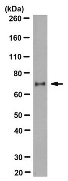
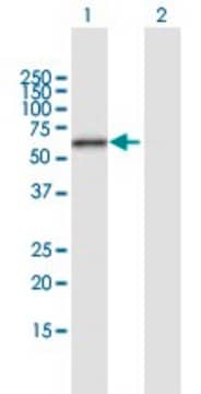
Western Blotting Analysis: 1 µg/mL from a representative lot detected Ero1 from 10 µg of the following cell lysates--A431, HEK293, HeLa, HepG2, human placenta tissue, HUVEC, jurkat, L6, NIH/3T3, PC12, PC3, and RAW264.7.

Immunoprecipitation Analysis: A representative lot immunoprecipitated Ero1 in Ero1 transfected HeLa cell lysate (Prof. R. Sitia, San Rafaele Scientific Institute, Milano, Italy).

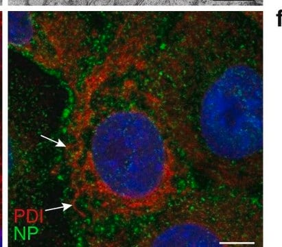
Immunofluorescence Analysis: A representative lot detected Ero1 in HeLa cell lysate (Prof. R. Sitia, San Rafaele Scientific Institute, Milano, Italy).

Quality

Evaluated by Western Blotting in HEK293 cell lysate.

Western Blotting Analysis: 1 µg/mL of this antibody detected Ero1 in 10 µg of HEK293 cell lysate.

Target description

~56 kDa observed. Uncharacterized band(s) may be observed in some cell lysates.

Physical form

Format: Purified
Protein G Purified
Purified mouse monoclonal IgG2aκ in buffer containing 0.1 M Tris-Glycine (pH 7.4), 150 mM NaCl with 0.05% sodium azide.

Storage and Stability

Stable for 1 year at 2-8°C from date of receipt.

Analysis Note

Control
HEK293 cell lysate

Other Notes

Concentration: Please refer to the Certificate of Analysis for the lot-specific concentration.

Disclaimer

Unless otherwise stated in our catalog or other company documentation accompanying the product(s), our products are intended for research use only and are not to be used for any other purpose, which includes but is not limited to, unauthorized commercial uses, in vitro diagnostic uses, ex vivo or in vivo therapeutic uses or any type of consumption or application to humans or animals.

Not finding the right product?  

Try our Product Selector Tool.

Storage Class Code

12 - Non Combustible Liquids

WGK

WGK 1

Flash Point(F)

Not applicable

Flash Point(C)

Not applicable


Certificates of Analysis (COA)

Search for Certificates of Analysis (COA) by entering the products Lot/Batch Number. Lot and Batch Numbers can be found on a product’s label following the words ‘Lot’ or ‘Batch’.

Already Own This Product?

Find documentation for the products that you have recently purchased in the Document Library.

Visit the Document Library

Samira Samtleben et al.
The European journal of neuroscience, 56(8), 5177-5190 (2022-09-10)
Multiple sclerosis (MS) and its animal models are characterized by cellular inflammation within the central nervous system (CNS). The sources and consequences of this inflammation are currently not completely understood. Critical signs and mediators of CNS inflammation are reactive oxygen
Shohei Fujii et al.
Proceedings of the National Academy of Sciences of the United States of America, 120(22), e2216857120-e2216857120 (2023-05-22)
Inositol 1,4,5-trisphosphate receptors (IP3Rs) are one of the two types of tetrameric ion channels that release calcium ion (Ca2+) from the endoplasmic reticulum (ER) into the cytosol. Ca2+ released via IP3Rs is a fundamental second messenger for numerous cell functions.
Jianchao Zhang et al.
The EMBO journal, 37(14) (2018-06-03)
Family with sequence similarity 20C (Fam20C), the physiological Golgi casein kinase, phosphorylates numerous secreted proteins that are involved in a wide variety of biological processes. However, the role of Fam20C in regulating proteins in the endoplasmic reticulum (ER) lumen is
Yu Yan et al.
Redox biology, 37, 101742-101742 (2020-10-13)
Hyperhomocysteinemia (HHcy) is related to liver diseases, such as nonalcoholic fatty liver (NAFL). Although the precise pathogenesis of NAFL is still largely unknown, the links between organs seem to play a vital role. The current study aimed to explore the
Na Zhang et al.
Atherosclerosis, 334, 39-47 (2021-09-04)
Hyperhomocysteinemia (HHcy) is an independent risk factor for atherosclerosis and plaque vulnerability. Macrophage apoptosis mediated by endoplasmic reticulum (ER) stress plays an important role in the pathogenesis of HHcy-aggravated atherosclerosis. Endoplasmic reticulum oxidoreductase 1α (Ero1α) is critical for ER stress-induced

Our team of scientists has experience in all areas of research including Life Science, Material Science, Chemical Synthesis, Chromatography, Analytical and many others.

Contact Technical Service