Accéder au contenu
Merck
Toutes les photos(5)

Documents

M2320

Sigma-Aldrich

Anti-MAP2 (2a+2b) antibody, Mouse monoclonal

~2 mg/mL, clone AP-20, purified from hybridoma cell culture

Synonyme(s) :

Anti-MAP2 (2a+2b) antibody, Mouse monoclonal, Anti-Microtubule Associated Protein 2

Se connecterpour consulter vos tarifs contractuels et ceux de votre entreprise/organisme


About This Item

Numéro MDL:
Code UNSPSC :
12352203
Nomenclature NACRES :
NA.41

Source biologique

mouse

Niveau de qualité

Conjugué

unconjugated

Forme d'anticorps

purified from hybridoma cell culture

Type de produit anticorps

primary antibodies

Clone

AP-20, monoclonal

Forme

buffered aqueous solution

Poids mol.

apparent mol wt 280 kDa

Espèces réactives

Xenopus, mouse, quail, human, bovine, rat, aquatic salamander

Conditionnement

antibody small pack of 25 μL

Concentration

~2 mg/mL

Technique(s)

immunocytochemistry: suitable
microarray: suitable
western blot: 1-3 μg/mL using rat brain preparation or rat cerebral cortex extract

Isotype

IgG1

Numéro d'accès UniProt

Conditions d'expédition

dry ice

Température de stockage

−20°C

Modification post-traductionnelle de la cible

unmodified

Informations sur le gène

human ... MAP2(4133)
mouse ... Mtap2(17756)
rat ... Map2(25595)

Vous recherchez des produits similaires ? Visite Guide de comparaison des produits

Description générale

MAP2 is the major microtubule associated protein of brain tissue. There are three forms of MAP2; two are similarily sized with apparent molecular weights of 280 kDa (MAP2a and MAP2b) and the third with a lower molecular weight of 70 kDa (MAP2c). In the newborn rat brain, MAP2b and MAP2c are present, while MAP2a is absent. Between postnatal days 10 and 20 MAP2a appears, at levels equal to that of MAP2b. At the same time, the level of MAP2c drops by 10-fold. This change happens during the period when dendrite growth is completed and when neurons have reached their mature morphology. MAP2 is degraded by a Cathepsin D like protease in the brain of aged rats. There is some indication that MAP2 is expressed at higher levels in some types of neurons than in others.
Monoclonal Anti-MAP2 (2a + 2b) (mouse IgG1 isotype) is derived from the hybridoma produced by the fusion of mouse myeloma cells and splenocytes from a mouse immunized with bovine microtubule associated protein 2 (MAP2).
Monoclonal Anti-MAP2 specificity was established by an immunoblot. The antibody localizes the high molecular weight forms of MAP2, namely MAP2a and MAP2b, but shows no reactivity with MAP2c. No crossreactivity is observed with MAP1, MAP5, tubulin, or tau (t).

Spécificité

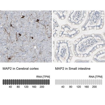
Monoclonal Anti-MAP2 antibody reacts with human, bovine, rat, mouse, Xenopus, salamander, and quail tissue or cells by immunocytochemical techniques utilizing either a fluorescent or peroxidase label, or by immunohistochemistry.

Immunogène

bovine MAP2

Application

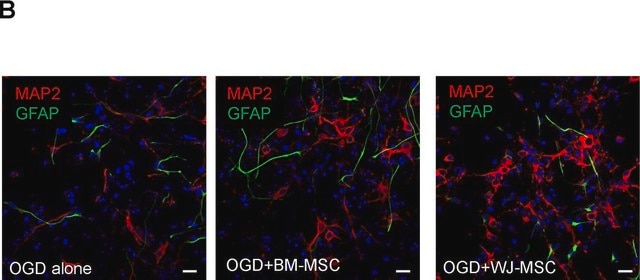
Monoclonal Anti-MAP2 (2a+2b) antibody produced in mouse has been used in immunocytochemistry.
Mouse monoclonal clone AP-20 anti-MAP2 (2a+2b) antibody is used to tag MAP2 for detection and quantitation by Western blotting and immunohistochemical (IHC) techniques. It is used as a probe to determine the roles of MAP2a and MAP2b in cell signaling especially in neural tissues.

Actions biochimiques/physiologiques

Microtubule associated protein 2 (MAP2) is known to promote microtubule assembly and to form side arms on microtubules. It also interacts with neurofilaments, actin and other elements of the cytoskeleton. MAP2 is degraded by a cathepsin D like protease in the brain of aged rats. There is some indication that MAP2 is expressed at higher levels in some types of neurons than in others. MAP2 is known to promote microtubule assembly and to form side-arms on microtubules. It also interacts with neurofilaments, actin and other elements of the cytoskeleton.

Forme physique

Solution in 0.01 M phosphate buffered saline, pH 7.4, containing 15 mM sodium azide.

Clause de non-responsabilité

Unless otherwise stated in our catalog or other company documentation accompanying the product(s), our products are intended for research use only and are not to be used for any other purpose, which includes but is not limited to, unauthorized commercial uses, in vitro diagnostic uses, ex vivo or in vivo therapeutic uses or any type of consumption or application to humans or animals.

Vous ne trouvez pas le bon produit ?  

Essayez notre Outil de sélection de produits.

Code de la classe de stockage

10 - Combustible liquids

Classe de danger pour l'eau (WGK)

WGK 2

Point d'éclair (°F)

Not applicable

Point d'éclair (°C)

Not applicable


Certificats d'analyse (COA)

Recherchez un Certificats d'analyse (COA) en saisissant le numéro de lot du produit. Les numéros de lot figurent sur l'étiquette du produit après les mots "Lot" ou "Batch".

Déjà en possession de ce produit ?

Retrouvez la documentation relative aux produits que vous avez récemment achetés dans la Bibliothèque de documents.

Consulter la Bibliothèque de documents

Antidepressant drugs diversely affect autophagy pathways in astrocytes and neurons?dissociation from cholesterol homeostasis
Zschocke J, et al.
Neuropsychopharmacology, 36(8), 1754-1754 (2011)
Shinya Hidano et al.
mBio, 7(6) (2016-11-12)
The local production of gamma interferon (IFN-γ) is important to control Toxoplasma gondii in the brain, but the basis for these protective effects is not fully understood. The studies presented here reveal that the ability of IFN-γ to inhibit parasite
The projection domain of MAP2b regulates microtubule protrusion and process formation in Sf9 cells
Belanger D, et al.
Journal of Cell Science, 115(7), 1523-1539 (2002)
Neurons Derived from Human Induced Pluripotent Stem Cells Integrate into Rat Brain Circuits and Maintain Both Excitatory and Inhibitory Synaptic Activities
Yin X, et al.
eNeuro, 6(4) (2019)
Carla M Cabral et al.
PLoS pathogens, 12(2), e1005447-e1005447 (2016-02-20)
Toxoplasma gondii, a common brain-tropic parasite, is capable of infecting most nucleated cells, including astrocytes and neurons, in vitro. Yet, in vivo, Toxoplasma is primarily found in neurons. In vitro data showing that interferon-γ-stimulated astrocytes, but not neurons, clear intracellular

Notre équipe de scientifiques dispose d'une expérience dans tous les secteurs de la recherche, notamment en sciences de la vie, science des matériaux, synthèse chimique, chromatographie, analyse et dans de nombreux autres domaines..

Contacter notre Service technique