Accéder au contenu
Merck
Toutes les photos(2)

Principaux documents

HPA012077

Sigma-Aldrich

Anti-FAM134B antibody produced in rabbit

Prestige Antibodies® Powered by Atlas Antibodies, affinity isolated antibody

Synonyme(s) :

FLJ20152, JK1

Se connecterpour consulter vos tarifs contractuels et ceux de votre entreprise/organisme


About This Item

Code UNSPSC :
12352203
Numéro HPA (Human Protein Atlas):
Nomenclature NACRES :
NA.41

Source biologique

rabbit

Niveau de qualité

Conjugué

unconjugated

Forme d'anticorps

affinity isolated antibody

Type de produit anticorps

primary antibodies

Clone

polyclonal

Gamme de produits

Prestige Antibodies® Powered by Atlas Antibodies

Forme

buffered aqueous glycerol solution

Espèces réactives

human

Technique(s)

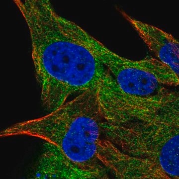
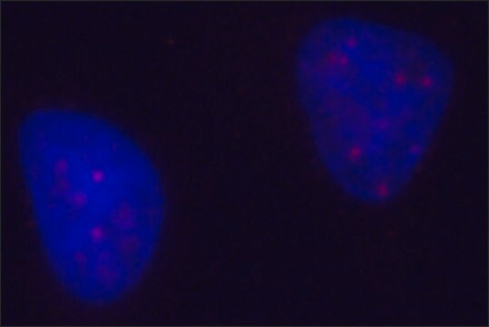
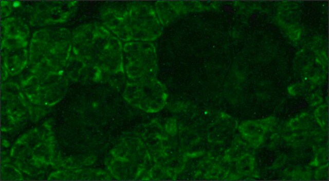
immunofluorescence: 0.25-2 μg/mL

Séquence immunogène

ENGMGTNDEDELSLGLPTELKRKKEQLDSGHRPSKETQSAAGLTLPLNSDQTFHLMSNLAGDVITAAVTAAIKDQLEGVQQALSQAAPIPEEDTDTEEGDDFELLDQSELDQIESELGLTQDQEAEAQQNKKSSG

Conditions d'expédition

wet ice

Température de stockage

−20°C

Modification post-traductionnelle de la cible

unmodified

Informations sur le gène

human ... FAM134B(54463)

Immunogène

family with sequence similarity 134, member B

Application

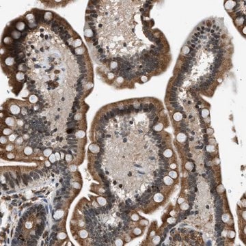
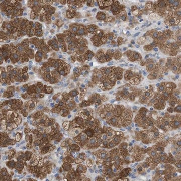
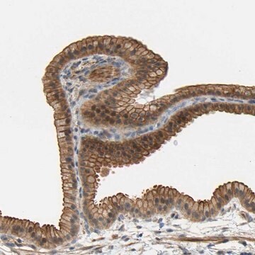
All Prestige Antibodies Powered by Atlas Antibodies are developed and validated by the Human Protein Atlas (HPA) project and as a result, are supported by the most extensive characterization in the industry.

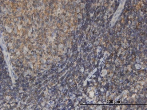
The Human Protein Atlas project can be subdivided into three efforts: Human Tissue Atlas, Cancer Atlas, and Human Cell Atlas. The antibodies that have been generated in support of the Tissue and Cancer Atlas projects have been tested by immunohistochemistry against hundreds of normal and disease tissues and through the recent efforts of the Human Cell Atlas project, many have been characterized by immunofluorescence to map the human proteome not only at the tissue level but now at the subcellular level. These images and the collection of this vast data set can be viewed on the Human Protein Atlas (HPA) site by clicking on the Image Gallery link. We also provide Prestige Antibodies® protocols and other useful information.

Caractéristiques et avantages

Prestige Antibodies® are highly characterized and extensively validated antibodies with the added benefit of all available characterization data for each target being accessible via the Human Protein Atlas portal linked just below the product name at the top of this page. The uniqueness and low cross-reactivity of the Prestige Antibodies® to other proteins are due to a thorough selection of antigen regions, affinity purification, and stringent selection. Prestige antigen controls are available for every corresponding Prestige Antibody and can be found in the linkage section.

Every Prestige Antibody is tested in the following ways:
  • IHC tissue array of 44 normal human tissues and 20 of the most common cancer type tissues.
  • Protein array of 364 human recombinant protein fragments.

Liaison

Corresponding Antigen APREST72121

Forme physique

Solution in phosphate buffered saline, pH 7.2, containing 40% glycerol and 0.02% sodium azide.

Informations légales

Prestige Antibodies is a registered trademark of Merck KGaA, Darmstadt, Germany

Vous ne trouvez pas le bon produit ?  

Essayez notre Outil de sélection de produits.

Code de la classe de stockage

10 - Combustible liquids

Classe de danger pour l'eau (WGK)

WGK 1

Point d'éclair (°F)

Not applicable

Point d'éclair (°C)

Not applicable


Faites votre choix parmi les versions les plus récentes :

Certificats d'analyse (COA)

Lot/Batch Number

Vous ne trouvez pas la bonne version ?

Si vous avez besoin d'une version particulière, vous pouvez rechercher un certificat spécifique par le numéro de lot.

Déjà en possession de ce produit ?

Retrouvez la documentation relative aux produits que vous avez récemment achetés dans la Bibliothèque de documents.

Consulter la Bibliothèque de documents

Rayene Berkane et al.
Nature communications, 14(1), 8364-8364 (2023-12-16)
Selective autophagy of the endoplasmic reticulum (ER), known as ER-phagy, is an important regulator of ER remodeling and essential to maintain cellular homeostasis during environmental changes. We recently showed that members of the FAM134 family play a critical role during
Umur Keles et al.
American journal of physiology. Gastrointestinal and liver physiology, 319(6), G733-G747 (2020-10-15)
Selective autophagy of the endoplasmic reticulum (ER), namely ER-phagy, is mediated by ER-localized receptors, which are recognized and sequestered by GABARAP/LC3B-decorated phagophores and transferred to lysosomes for degradation. Being one such receptor, FAM134B plays critical roles in cellular processes such
Francescopaolo Iavarone et al.
EMBO reports, 25(8), 3651-3677 (2024-07-23)
Endoplasmic reticulum (ER) remodeling is vital for cellular organization. ER-phagy, a selective autophagy targeting ER, plays an important role in maintaining ER morphology and function. The FAM134 protein family, including FAM134A, FAM134B, and FAM134C, mediates ER-phagy. While FAM134B mutations are
Laura Cinque et al.
The EMBO journal, 39(17), e105696-e105696 (2020-07-28)
Lysosomal degradation of the endoplasmic reticulum (ER) via autophagy (ER-phagy) is emerging as a critical regulator of cell homeostasis and function. The recent identification of ER-phagy receptors has shed light on the molecular mechanisms underlining this process. However, the signaling
Alison Forrester et al.
The EMBO journal, 38(2) (2018-12-19)
Autophagy is a cytosolic quality control process that recognizes substrates through receptor-mediated mechanisms. Procollagens, the most abundant gene products in Metazoa, are synthesized in the endoplasmic reticulum (ER), and a fraction that fails to attain the native structure is cleared

Notre équipe de scientifiques dispose d'une expérience dans tous les secteurs de la recherche, notamment en sciences de la vie, science des matériaux, synthèse chimique, chromatographie, analyse et dans de nombreux autres domaines..

Contacter notre Service technique