Accéder au contenu
Merck
Toutes les photos(7)

Documents

A2103

Sigma-Aldrich

Anti-Actin, N-terminal antibody produced in rabbit

enhanced validation

~0.5 mg/mL, affinity isolated antibody, buffered aqueous solution

Synonyme(s) :

Actin Detection Antibody, Rabbit Anti-Actin

Se connecterpour consulter vos tarifs contractuels et ceux de votre entreprise/organisme


About This Item

Numéro MDL:
Code UNSPSC :
12352203
Nomenclature NACRES :
NA.41

Source biologique

rabbit

Niveau de qualité

Conjugué

unconjugated

Forme d'anticorps

affinity isolated antibody

Type de produit anticorps

primary antibodies

Clone

polyclonal

Forme

buffered aqueous solution

Poids mol.

antigen 42 kDa

Espèces réactives

frog, rat, mouse, chicken, human

Conditionnement

antibody small pack of 25 μL

Validation améliorée

independent
Learn more about Antibody Enhanced Validation

Concentration

~0.5 mg/mL

Technique(s)

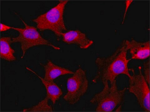
immunocytochemistry: 1-2 μg/mL using cultured chicken fibroblasts
immunohistochemistry (formalin-fixed, paraffin-embedded sections): 2-4 μg/mL using sections of human appendix, mouse heart, and frog skeletal muscle
indirect immunofluorescence: suitable
western blot: 0.5-1 μg/mL using whole extract of the human epitheloid carcinoma HeLa cell line.
western blot: 2-4 ng/mL using whole extract of rat skeletal muscle

Numéro d'accès UniProt

Conditions d'expédition

wet ice

Température de stockage

−20°C

Modification post-traductionnelle de la cible

unmodified

Description générale

Actin is a cytoskeletal protein that regulates cell motility, secretion, phagocytosis and cytokinesis. The NH2-terminal of actin may function as an antigen. This terminal may also modulate actin interactions and may associate with proteins such as myosin.
Actin is one of the most conserved eukaryotic proteins, which is expressed in mammals and birds as at least six major different isoforms. Four of them represent the differentiation markers of muscle tissues and two are found practically in all non-muscle cells. Actin isoforms show >90% overall sequence homology, but only 50-60% homology in their 18 N-terminal residues. Actin is present in cells both as a globular monomer (G-actin) and as a polymer in filamentous actin (F-actin) that participates in the formation of a variety of stable and labile structures. The presence of actin in cell nuclei and cell membranes has been reported.

Immunogène

synthetic actin N-terminal nonapeptide conjugated to KLH.

Application

Anti-Actin, N-terminal antibody produced in rabbit has been used in
  • immunoblotting
  • quantitative chromatin immunoprecipitation (qChIP)

Protein lysates generated from embryonic mouse limb tissue was analyzed by western blot using actin as the loading control. Actin was detected using rabbit anti-actin at 1:8000. Actin detected by a rabbit anti-actin antibody was used as a loading control for MCF7 cell lysates . Actin was detected at 42 kDa.
Rabbit anti-actin, N-terminal antibody can be used for western blotting assays. The antibody can also be used for immunocytochemistry at 1-2μg/mL using cultured chicken fibroblasts. Furthermore, the antibody is suitable for use in immunohistochemistry at a concentration of 2-4μg/mL using sections of human appendix, mouse heart, and frog skeletal muscle.

Actions biochimiques/physiologiques

Actin and myosin are constituents of many cell types and are involved in a myriad of cellular processes including maintenance of cell shape, locomotion, secretion, cytoplasmic streaming, phagocytosis, and cytokinesis. The N-terminal domain of actin appears to be a major antigenic region of the molecule. Different isoforms can coexist in the same cell but are differentially regulated, and in most cases cannot substitute for each other functionally.

Forme physique

Solution in 0.01 M phosphate buffered saline, pH 7.4, containing 1% BSA and 15 mM sodium azide.

Autres remarques

To view an Actin antibody selection guide, please visit www.sigmaaldrich.com/actin.

Clause de non-responsabilité

Unless otherwise stated in our catalog or other company documentation accompanying the product(s), our products are intended for research use only and are not to be used for any other purpose, which includes but is not limited to, unauthorized commercial uses, in vitro diagnostic uses, ex vivo or in vivo therapeutic uses or any type of consumption or application to humans or animals.

Vous ne trouvez pas le bon produit ?  

Essayez notre Outil de sélection de produits.

Code de la classe de stockage

12 - Non Combustible Liquids

Classe de danger pour l'eau (WGK)

WGK 2

Équipement de protection individuelle

Eyeshields, Gloves, multi-purpose combination respirator cartridge (US)


Certificats d'analyse (COA)

Recherchez un Certificats d'analyse (COA) en saisissant le numéro de lot du produit. Les numéros de lot figurent sur l'étiquette du produit après les mots "Lot" ou "Batch".

Déjà en possession de ce produit ?

Retrouvez la documentation relative aux produits que vous avez récemment achetés dans la Bibliothèque de documents.

Consulter la Bibliothèque de documents

Nuclear actin polymerization is required for transcriptional reprogramming of Oct4 by oocytes
Miyamoto K, et al.
Genes & Development, 25(9), 946-958 (2011)
Monoclonal antibodies against muscle actin isoforms: epitope identification and analysis of isoform expression by immunoblot and immunostaining in normal and regenerating skeletal muscle
Chaponnier C and Gabbiani G
F1000Research, 5 (2016)
Kristine Raaby Jakobsen et al.
Journal of molecular signaling, 8(1), 9-9 (2013-09-06)
Protrusions of cancer cells conferrers a vital function for cell migration and metastasis. Protein and RNA localization mechanisms have been extensively examined and shown to play pivotal roles for the functional presence of specific protein components in cancer cell protrusions.
Luciferase expression and bioluminescence does not affect tumor cell growth in vitro or in vivo
Tiffen JC, et al.
Molecular Cancer, 9(1), 299-299 (2010)
Kale S Bongers et al.
American journal of physiology. Endocrinology and metabolism, 305(7), E907-E915 (2013-08-15)
Skeletal muscle denervation causes muscle atrophy via complex molecular mechanisms that are not well understood. To better understand these mechanisms, we investigated how muscle denervation increases growth arrest and DNA damage-inducible 45α (Gadd45a) mRNA in skeletal muscle. Previous studies established

Notre équipe de scientifiques dispose d'une expérience dans tous les secteurs de la recherche, notamment en sciences de la vie, science des matériaux, synthèse chimique, chromatographie, analyse et dans de nombreux autres domaines..

Contacter notre Service technique