Accéder au contenu
Merck
Toutes les photos(2)

Key Documents

MABT824

Sigma-Aldrich

Anti-gamma-Actin/ACTG1 Antibody, clone 2A3

clone 2A3, from mouse

Synonyme(s) :

Actin, cytoplasmic 2, gamma-Actin

Se connecterpour consulter vos tarifs contractuels et ceux de votre entreprise/organisme


About This Item

Code UNSPSC :
12352203
eCl@ss :
32160702
Nomenclature NACRES :
NA.41

Source biologique

mouse

Niveau de qualité

Forme d'anticorps

purified immunoglobulin

Type de produit anticorps

primary antibodies

Clone

2A3, monoclonal

Espèces réactives

mouse, rat, human, chicken

Réactivité de l'espèce (prédite par homologie)

mammals (based on 100% sequence homology)

Technique(s)

immunocytochemistry: suitable
western blot: suitable

Isotype

IgG2bκ

Numéro d'accès NCBI

Numéro d'accès UniProt

Conditions d'expédition

wet ice

Modification post-traductionnelle de la cible

unmodified

Informations sur le gène

human ... ACTG1(71)

Description générale

Actin, cytoplasmic 2 (UniProt P63261; also known as Gamma-actin) is encoded by the ACTG1 (also known as ACTG, BRWS2, DFNA20, DFNA26) gene (Gene ID 71) in human. Actins are globular multi-functional proteins that serve as the basic building blocks of cytoskeletal microfilaments and are among the most conserved eukaryotic proteins. Six actin types exist, skeletal muscle alpha-actin is encoded by the ACTA1 gene, smooth muscle alpha-actin by the ACTA2 gene, cytoplasmic beta-actin by the ACTB gene, cardiac muscle alpha-actin by the ACTC1 gene, cytoplasmic gamma-actin by the ACTG1 gene, and smooth muscle gamma-actin by the ACTG2 (a.k.a. ACTA3) gene. Although actins show >90% overall sequence homology, isoforms do show spatial, temporal, and tissue-specific expression patterns and only 50-60% homology is found in their 18 N-terminal residues. Cytoplasmic β and γ-actins, are thought to be present in all cells, while the other four actin isoforms are typically found in specific adult muscle tissue types. Actins exist in a variety of structural states, depending on the specific ionic conditions or the interaction with ligand proteins. The oligomeric and polymeric forms that actin molecules assume are dependent on the distinct conformations they adopt.

Spécificité

Clone 2A3 detected BSA conjugated with cytoplasmic gamma-actin N-terminal peptide, but not BSA conjugated with N-terminal peptides derived from the other 5 actin types (Dugina, V., et al. (2009). J. Cell Sci. 122(Pt 16):2980-2988).

Immunogène

Epitope: Near N-terminus.
KLH-conjugated linear peptide corresponding to the the N-terminal sequence of human gamma-Actin/ACTG1.

Application

Research Category
Cell Structure
Research Sub Category
Cytoskeleton
This Anti-gamma-Actin/ACTG1 Antibody, clone 2A3 is validated for use in Western Blotting, Immunocytochemistry for the detection of ACTG1.
Western Blotting Analysis: 0.5 µg/mL from a representative lot detected gamma-Actin/ACTG1 in 10 µg of NIH/3T3 cell lysate.
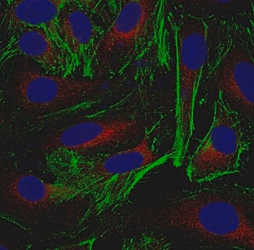
Immunocytochemistry Analysis: 5.0 µg/mL from a representative lot detected gamma-Actin/ACTG1 in HUVECs, HeLa and A431 cells.
Western Blotting Analysis: A representative lot detected downregulated cytoplasmic gamma-actin levels in stimulated (by A23187, TRAP-6, TNF, LPS, or IFN-γ) human cerebral microvascular endothelial D3 cells (hCMEC/D3) and their microparticles (MPs) when compared with unstimulated hCMEC/D3 and their MPs (Latham, S.L., et al. (2013). FASEB J. 27(2):672-683).
Western Blotting Analysis: A representative lot detected siRNA-mediated downregulation of cytoplasmic gamma-actin in A549 human lung carcinoma cells (Miazza, V., et al. (2011). Virology. 410(1):7-16).
Western Blotting Analysis: A representative lot detected cytoplasmic gamma-actin, but not beta-actin separated by 2-D gel electrophoresis of purified chicken gizzard actins or total protein extracts from human subcutaneous fibroblasts (HSCFs), canine MDCK cells, and rat aorta tissue (Dugina, V., et al. (2009). J. Cell Sci. 122(Pt 16):2980-2988).
Western Blotting Analysis: A representative lot detected BSA conjugated with cytoplasmic gamma-actin N-terminal peptide, but not BSA conjugated with N-terminal peptides derived from the 5 other actin types (Dugina, V., et al. (2009). J. Cell Sci. 122(Pt 16):2980-2988).
Immunocytochemistry Analysis: A representative lot detected TNF-stimulated localization of γ-actin into thick, intensely staining stress fibers concentrated apically in a submembranous network in human cerebral microvascular endothelial D3 cells (hCMEC/D3) (Latham, S.L., et al. (2013). FASEB J. 27(2):672-683).
Immunocytochemistry Analysis: A representative lot detected similar γ-actin cytoskeleton structure in human cerebral microvascular endothelial D3 cells (hCMEC/D3) with or without Rho kinase inhibitor Y-27632 treatment (Latham, S.L., et al. (2013). FASEB J. 27(2):672-683).
Immunocytochemistry Analysis: A representative lot detected a drastic subcellular redistribution of gamma-actin following Sendai virus infection of polarized Madin-Darby canine kidney (MDCK) epithelial cells by fluorescent immunocytochemistry staining of paraformaldehyde-fixed, methanol-treated cells (Miazza, V., et al. (2011). Virology. 410(1):7-16).
Immunocytochemistry Analysis: A representative lot detected cytoplasmic gamma-actin subcellular localization distinct from that of beta-actin in both spreading and stationary cells by fluorescent immunocytochemistry, using paraformaldehyde-fixed, methanol-treated HSCF human subcutaneous fibroblasts, HaCaT human keratinocytes, WI38 human embryonic fibroblasts,and Madin-Darby canine kidney (MDCK) cells (Dugina, V., et al. (2009). J. Cell Sci. 122(Pt 16):2980-2988).

Qualité

Evaluated by Western Blotting in HeLa cell lysate.

Western Blotting Analysis: 0.5 µg/mL of this antibody detected gamma-Actin/ACTG1 in 10 µg of HeLa cell lysate.

Description de la cible

~39-45 kDa observed. Uncharacterized band(s) may appear in some lysates.

Forme physique

Format: Purified
Protein G Purified
Purified mouse monoclonal IgG2bκ antibody in buffer containing 0.1 M Tris-Glycine (pH 7.4), 150 mM NaCl with 0.05% sodium azide.

Stockage et stabilité

Stable for 1 year at 2-8°C from date of receipt.

Autres remarques

Concentration: Please refer to lot specific datasheet.

Clause de non-responsabilité

Unless otherwise stated in our catalog or other company documentation accompanying the product(s), our products are intended for research use only and are not to be used for any other purpose, which includes but is not limited to, unauthorized commercial uses, in vitro diagnostic uses, ex vivo or in vivo therapeutic uses or any type of consumption or application to humans or animals.

Vous ne trouvez pas le bon produit ?  

Essayez notre Outil de sélection de produits.

Code de la classe de stockage

12 - Non Combustible Liquids

Classe de danger pour l'eau (WGK)

WGK 1

Point d'éclair (°F)

Not applicable

Point d'éclair (°C)

Not applicable


Certificats d'analyse (COA)

Recherchez un Certificats d'analyse (COA) en saisissant le numéro de lot du produit. Les numéros de lot figurent sur l'étiquette du produit après les mots "Lot" ou "Batch".

Déjà en possession de ce produit ?

Retrouvez la documentation relative aux produits que vous avez récemment achetés dans la Bibliothèque de documents.

Consulter la Bibliothèque de documents

Tomoyuki Hatano et al.
Journal of cell science, 131(8) (2018-03-15)
Actins are major eukaryotic cytoskeletal proteins, and they are involved in many important cell functions, including cell division, cell polarity, wound healing and muscle contraction. Despite obvious drawbacks, muscle actin, which is easily purified, is used extensively for biochemical studies
Chengqing Qu et al.
Journal of the American Society of Nephrology : JASN, 33(1), 155-173 (2021-11-12)
Actin stress fibers are abundant in cultured cells, but little is known about them in vivo. In podocytes, much evidence suggests that mechanobiologic mechanisms underlie podocyte shape and adhesion in health and in injury, with structural changes to actin stress
Rikki M Garner et al.
Cytoskeleton (Hoboken, N.J.), 77(5-6), 181-196 (2020-02-20)
Observations of actin dynamics in living cells using fluorescence microscopy have been foundational in the exploration of the mechanisms underlying cell migration. We used CRISPR/Cas9 gene editing to generate neutrophil-like HL-60 cell lines expressing GFP-β-actin from the endogenous locus (ACTB).
Christine Chaponnier et al.
F1000Research, 5, 416-416 (2016-06-24)
Higher vertebrates (mammals and birds) express six different highly conserved actin isoforms that can be classified in three subgroups: 1) sarcomeric actins, α-skeletal (α-SKA) and α-cardiac (α-CAA), 2) smooth muscle actins (SMAs), α-SMA and γ-SMA, and 3) cytoplasmic actins (CYAs)
Kristen Skruber et al.
Current biology : CB, 30(14), 2651-2664 (2020-05-30)
Cells have many types of actin structures, which must assemble from a common monomer pool. Yet, it remains poorly understood how monomers are distributed to and shared between different filament networks. Simplified model systems suggest that monomers are limited and

Notre équipe de scientifiques dispose d'une expérience dans tous les secteurs de la recherche, notamment en sciences de la vie, science des matériaux, synthèse chimique, chromatographie, analyse et dans de nombreux autres domaines..

Contacter notre Service technique