Accéder au contenu
Merck
Toutes les photos(3)

Documents

MAB5258

Sigma-Aldrich

Anti-Synaptophysin Antibody, clone SY38

clone SY38, Chemicon®, from mouse

Synonyme(s) :

synaptophysin, Synaptophysin

Se connecterpour consulter vos tarifs contractuels et ceux de votre entreprise/organisme


About This Item

Code UNSPSC :
12352203
eCl@ss :
32160702
Nomenclature NACRES :
NA.41

Source biologique

mouse

Niveau de qualité

Forme d'anticorps

purified immunoglobulin

Type de produit anticorps

primary antibodies

Clone

SY38, monoclonal

Espèces réactives

bovine, rat, avian, fish, mouse, human

Fabricant/nom de marque

Chemicon®

Technique(s)

flow cytometry: suitable
immunocytochemistry: suitable
immunohistochemistry: suitable
western blot: suitable

Isotype

IgG1κ

Numéro d'accès NCBI

Numéro d'accès UniProt

Conditions d'expédition

wet ice

Modification post-traductionnelle de la cible

unmodified

Informations sur le gène

human ... SYP(6855)

Description générale

Synaptophysin is a 38 kDa glycoprotein present in the membrane of neuronal presynaptic vesicles in brain, spinal cord, retina, vesicles of adrenal medulla, neuromuscular junctions, and endocrine cells. It is also expressed by neuroendocrine cells throughout the body, both normal and neoplastic. Synaptophysin is a useful marker for the identification of normal neuroendocrine cells and neuroendocrine neoplasms. This antibody reacts with neuroendocrine cells of human adrenal medulla, carotid body, skin, pituitary gland, thyroid, lung, pancreas, and gastointestinal mucosa. It also reacts with a wide spectrum of neuroendocrine neoplasms of neural type including neuroblastomas, ganglioneuroblastomas, ganglioneuromas, pheochromocytomas, chromaffin, and non chromaffin paragangliomas.

Spécificité

The antibody reacts with presynaptic vesicles of cerebral and spinal neurons, of neuromuscular endplates and with the retina of man, cow, rat and mouse. The antibody also reacts with vesicles of adrenal medulla and islet cells, and additionally allows specific staining of neuronal, adrenal and neuroepithelial tumors, such as pheochromocytoma, paraganglioma, islet cell tumors (incl. Insulinoma), medullary thyroid carcinoma and diverse pulmonary and gastro-intestinal carcinoids. The antibody also stains neurosecretory vesicles of certain culture cells, e.g. of the rat cell line PC-12.

Immunogène

Vesicular fraction of bovine brain. The SY38 epitope is to a pentapeptide repeat structure in the carboxy-terminal cytoplasmic tail of synaptophysin (Knaus & Betz 1990).

Application

Detect Synaptophysin using this Anti-Synaptophysin Antibody, clone SY38 validated for use in FC, IC, IH & WB.
Flow Cytometry (FACS):
A previous lot of this antibody was used in Flow Cytometry.

Western Blot:
(Provoda, 2000) Reacts with a 38 kDa transmembrane glycoprotein

Immunohistochemistry:
For relatively cytoplasm-rich neuroendocrinic tumors a final concentration of 1 µg/mL is recommended. Concerning cytoplasm deficient tumors a concentration of 2 µg/mL should be used. Ideal frozen sections (4-5 mm) are obtained from shock-frozen tissue samples. The frozen sections are air-dried and then fixed with acetone for 5-10 min at -20°C. Excess acetone is allowed to evaporate at room temperature. Material fixed in alcohol or formalin and embedded in paraffin can also be used.
It is advantageous to block unspecific binding sites by overlaying the sections with fetal calf serum for 20-30 min at room temperature. Excess of fetal calf serum is removed by decanting before application of the anti-body solution.
Cytocentrifuge preparations of single cells or cell smears are also fixed in acetone. These preparations should but should be followed directly by antibody treatment.

Optimal working dilutions must be determined by end user.
Research Category
Neuroscience
Research Sub Category
Synapse & Synaptic Biology

Qualité

Routinely evaluated by Western Blot on mouse brain lysates.

Western Blot Analysis:
1:1000 dilution of this lot detected synaptophysin on 10 μg of mouse brain lysates.

Description de la cible

38 kDa

Liaison

Replaces: 04-1019

Forme physique

Format: Purified
Protein A purified
Purified mouse monoclonal IgG1κ in buffer containing 20 mM sodium phosphate buffer, 0.25 M NaCl, containing 0.1% sodium azide.

Stockage et stabilité

Stable for 6 months at 2-8ºC from date of receipt. DO NOT FREEZE.

Remarque sur l'analyse

Control
Positive Control: Pancreas tissue
Negative Control: Normal mouse serum
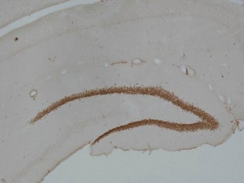
Rat hippocampus tissue, mouse brain lysate.

Autres remarques

Concentration: Please refer to the Certificate of Analysis for the lot-specific concentration.

Informations légales

CHEMICON is a registered trademark of Merck KGaA, Darmstadt, Germany

Clause de non-responsabilité

Unless otherwise stated in our catalog or other company documentation accompanying the product(s), our products are intended for research use only and are not to be used for any other purpose, which includes but is not limited to, unauthorized commercial uses, in vitro diagnostic uses, ex vivo or in vivo therapeutic uses or any type of consumption or application to humans or animals.

Vous ne trouvez pas le bon produit ?  

Essayez notre Outil de sélection de produits.

Code de la classe de stockage

10 - Combustible liquids

Classe de danger pour l'eau (WGK)

WGK 2

Point d'éclair (°F)

Not applicable

Point d'éclair (°C)

Not applicable


Certificats d'analyse (COA)

Recherchez un Certificats d'analyse (COA) en saisissant le numéro de lot du produit. Les numéros de lot figurent sur l'étiquette du produit après les mots "Lot" ou "Batch".

Déjà en possession de ce produit ?

Retrouvez la documentation relative aux produits que vous avez récemment achetés dans la Bibliothèque de documents.

Consulter la Bibliothèque de documents

Claudia Fuchs et al.
Neurobiology of disease, 70, 53-68 (2014-06-22)
Mutations in the X-linked cyclin-dependent kinase-like 5 (CDKL5) gene have been identified in a neurodevelopmental disorder characterized by early-onset intractable seizures, severe developmental delay, intellectual disability, and Rett's syndrome-like features. Since the physiological functions of CDKL5 still need to be
Iwona Ksiazek et al.
The Journal of neuroscience : the official journal of the Society for Neuroscience, 27(27), 7183-7195 (2007-07-06)
Agrin-deficient mice die at birth because of aberrant development of the neuromuscular junctions. Here, we examined the role of agrin at brain synapses. We show that agrin is associated with excitatory but not inhibitory synapses in the cerebral cortex. Most
Hengchang Li et al.
Frontiers in neuroscience, 13, 706-706 (2019-07-30)
Children, after multiple exposures to general anesthesia, appear to be at an increased risk of developing learning disabilities. Almost all general anesthetics-including sevoflurane, which is commonly used for children-are potentially neurotoxic to the developing brain. Anesthesia exposure during development might
Sven Akkerman et al.
PloS one, 9(11), e111692-e111692 (2014-11-06)
Drug effects are usually evaluated in animals housed under maximally standardized conditions. However, it is assumed that an enriched environment (EE) more closely resembles human conditions as compared to maximally standardized laboratory conditions. In the present study, we examined the
Arielle R Strzelewicz et al.
Hormones and behavior, 111, 46-59 (2019-02-02)
Early life exposure to a low security setting, characterized by a scarcity of resources and limited food access, increases the risk for psychiatric illness and metabolic dysfunction. We utilized a translational rat model to mimic a low security environment and

Notre équipe de scientifiques dispose d'une expérience dans tous les secteurs de la recherche, notamment en sciences de la vie, science des matériaux, synthèse chimique, chromatographie, analyse et dans de nombreux autres domaines..

Contacter notre Service technique