Skip to Content
Merck
All Photos(1)

Key Documents

P6248

Sigma-Aldrich

Anti-Parkin antibody, Mouse monoclonal

clone PRK8, purified from hybridoma cell culture

Synonym(s):

Anti-AR-JP, Anti-LPRS2, Anti-PARK2, Anti-PDJ

Sign Into View Organizational & Contract Pricing


About This Item

MDL number:
UNSPSC Code:
12352203
NACRES:
NA.41

biological source

mouse

Quality Level

conjugate

unconjugated

antibody form

purified immunoglobulin

antibody product type

primary antibodies

clone

PRK8, monoclonal

form

buffered aqueous solution

species reactivity

mouse, rat, hamster, human

concentration

~2 mg/mL

technique(s)

microarray: suitable
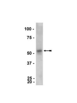
western blot: 0.25-0.5 μg/mL using using rat brain cytosolic S1 extract

isotype

IgG2b

shipped in

dry ice

storage temp.

−20°C

target post-translational modification

unmodified

Gene Information

human ... PARK2(5071)
mouse ... Park2(50873)
rat ... Park2(56816)

General description

Monoclonal Anti- Parkin (mouse IgG2b isotype) is derived from the hybridoma PRK8 produced by the fusion of mouse myeloma cells (SP2 cells) and splenocytes from BALB/c mice immunized with recombinant human Parkin. Parkin is an ubiquitin-protein ligase expresses in many tissues such as brain, testis, skeletal muscle and heart. The parkin gene encodes a protein of 465 amino acid residues with moderate similarity to ubiquitin at the amino-terminus and a ring in between ring fingers (IBR)-ring-finger motif at the carboxyl-terminus. The gene has 12 exons.
Parkin is an ubiquitin-protein ligase expresses in many tissues such as brain, testis, skeletal muscle and heart. It is a RING-type E3 ubiquitin-protein ligase that facilitates the ubiquitination of misfolded proteins and protects from neurotoxic effect caused by these proteins. Monoclonal anti-parkin antibody can be used in microarray and immunoprecipitation assays. It can also be used in western blotting. Mouse anti-parkin antibody reacts specifically with parkin (50kD approx) of rat, hamster, mouse and human.

Immunogen

recombinant human Parkin.

Application

Monoclonal anti-parkin antibody has been used in:
  • Enzyme linked immunosorbent assay (ELISA)
  • Immunoblotting
  • Immunoprecipitation[63}
  • Immunocytochemistry
  • to label parkin in pull-down assay

Physical form

Solution in 0.01 M phosphate buffered saline, pH 7.4, containing 15 mM sodium azide.

Disclaimer

Unless otherwise stated in our catalog or other company documentation accompanying the product(s), our products are intended for research use only and are not to be used for any other purpose, which includes but is not limited to, unauthorized commercial uses, in vitro diagnostic uses, ex vivo or in vivo therapeutic uses or any type of consumption or application to humans or animals.

Not finding the right product?  

Try our Product Selector Tool.

Storage Class Code

10 - Combustible liquids

WGK

WGK 3

Flash Point(F)

Not applicable

Flash Point(C)

Not applicable

Personal Protective Equipment

dust mask type N95 (US), Eyeshields, Gloves

Certificates of Analysis (COA)

Search for Certificates of Analysis (COA) by entering the products Lot/Batch Number. Lot and Batch Numbers can be found on a product’s label following the words ‘Lot’ or ‘Batch’.

Already Own This Product?

Find documentation for the products that you have recently purchased in the Document Library.

Visit the Document Library

Koji Yamano et al.
The Journal of biological chemistry, 290(42), 25199-25211 (2015-08-12)
Damaged mitochondria are eliminated through autophagy machinery. A cytosolic E3 ubiquitin ligase Parkin, a gene product mutated in familial Parkinsonism, is essential for this pathway. Recent progress has revealed that phosphorylation of both Parkin and ubiquitin at Ser(65) by PINK1
Victoriane Peugnet et al.
Antioxidants (Basel, Switzerland), 11(4) (2022-04-24)
Heart failure, mostly associated with cardiac hypertrophy, is a major cause of illness and death. Oxidative stress causes accumulation of reactive oxygen species (ROS), leading to mitochondrial dysfunction, suggesting that mitochondria-targeted therapies could be effective in this context. The purpose
Genomic organization and expression of parkin in Drosophila melanogaster
Bae JY, et al.
Experimental & Molecular Medicine, 35(5), 393-393 (2003)
Parkin regulates kainate receptors by interacting with the GluK2 subunit
Maraschi AM, et al,
Nature Communications, 5(46) (2014)
Aaron C Pawlyk et al.
The Journal of biological chemistry, 278(48), 48120-48128 (2003-09-16)
Autosomal recessive juvenile parkinsonism is a movement disorder associated with the degeneration of dopaminergic neurons in substantia nigra pars compacta. The loss of functional parkin caused by parkin gene mutations is the most common single cause of juvenile parkinsonism. Parkin

Our team of scientists has experience in all areas of research including Life Science, Material Science, Chemical Synthesis, Chromatography, Analytical and many others.

Contact Technical Service