Skip to Content
Merck
All Photos(2)

Key Documents

M8064

Sigma-Aldrich

Anti-Myosin IIA, non muscle antibody produced in rabbit

affinity isolated antibody, buffered aqueous solution

Synonym(s):

Anti-BDPLT6, Anti-DFNA17, Anti-EPSTS, Anti-FTNS, Anti-MATINS, Anti-MHA, Anti-NMHC-II-A, Anti-NMMHC-IIA, Anti-NMMHCA

Sign Into View Organizational & Contract Pricing


About This Item

MDL number:
UNSPSC Code:
12352203
NACRES:
NA.41

biological source

rabbit

Quality Level

conjugate

unconjugated

antibody form

affinity isolated antibody

antibody product type

primary antibodies

clone

polyclonal

form

buffered aqueous solution

mol wt

antigen ~200 kDa

species reactivity

rat, human, canine

packaging

antibody small pack of 25 μL

technique(s)

indirect immunofluorescence: 1:100 using cultured rat NRK cells
microarray: suitable
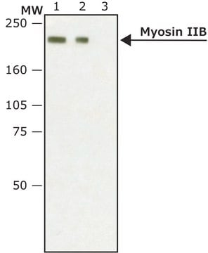
western blot: 1:1,000 using whole cell extracts of cultured dog MDCK kidney cells and cultured human Jurkat cells

UniProt accession no.

shipped in

dry ice

storage temp.

−20°C

target post-translational modification

unmodified

Gene Information

human ... MYH9(4627)
rat ... Myh9(25745)

General description

Myosin heavy chain 9 (MYH9) gene codes for the nonmuscle myosin heavy chain IIA.
Myosin heavy chain 9 is expressed in platelets and the gene encoding it is localized on chromosome 22.

Immunogen

synthetic peptide corresponding to amino acids 1949-1960 of human nonmuscle myosin IIA.

Application

Anti-Myosin IIA, non muscle antibody produced in rabbit has been used in:
  • immunofluorescence
  • immunohistochemistry
  • fixed cell staining/ immunofluorescence staining
  • immunoblot
  • immunoprecipitation

Biochem/physiol Actions

Nonmuscle myosin II is involved in cell motility and adhesion, cytokinesis, vesicular transport, intracellular force generation and in morphogenesis during development. Its activity is regulated by light chain and possibly heavy chain phosphorylation and by association with proteins such as Mts1. Mutations in the NMHCA gene are found in several syndromes associated with megakaryocyte/platelet/leukocyte disorders. Mutations in the MYH9 gene causes a spectrum of macrothrombocytopenia disorders with neutrophil inclusions, termed as MYH9 disorders.

Physical form

Solution in 0.01 M phosphate buffered saline, pH 7.4, containing 1% BSA and 15 mM sodium azide.

Disclaimer

Unless otherwise stated in our catalog or other company documentation accompanying the product(s), our products are intended for research use only and are not to be used for any other purpose, which includes but is not limited to, unauthorized commercial uses, in vitro diagnostic uses, ex vivo or in vivo therapeutic uses or any type of consumption or application to humans or animals.

Not finding the right product?  

Try our Product Selector Tool.

Storage Class Code

12 - Non Combustible Liquids

WGK

nwg

Flash Point(F)

Not applicable

Flash Point(C)

Not applicable


Certificates of Analysis (COA)

Search for Certificates of Analysis (COA) by entering the products Lot/Batch Number. Lot and Batch Numbers can be found on a product’s label following the words ‘Lot’ or ‘Batch’.

Already Own This Product?

Find documentation for the products that you have recently purchased in the Document Library.

Visit the Document Library

The role of stress fibers in the shape determination mechanism of fish keratocytes
Nakata T, et al.
Biophysical Journal, 110(2), 481-492 (2016)
Hemostasis and Thrombosis
Hematopathology, 57-105 (2018)
Nonmuscle myosin-2: mix and match
Heissler SM and Manstein DJ
Cellular and Molecular Life Sciences, 70(1), 1-21 (2013)
Y Chen et al.
Journal of thrombosis and haemostasis : JTH, 11(12), 2163-2175 (2013-10-30)
Mutations in the MYH9 gene cause autosomal dominant MYH9-related diseases (MYH9-RD) that associate macrothrombocytopenia with various other clinical conditions. The mechanisms giving rise to giant platelets remain poorly understood. To study the proplatelet formation (PPF) derived from megakaryocytes (MKs) generated
Praveen Suraneni et al.
Molecular biology of the cell, 26(5), 901-912 (2015-01-09)
Cells employ protrusive leading edges to navigate and promote their migration in diverse physiological environments. Classical models of leading-edge protrusion rely on a treadmilling dendritic actin network that undergoes continuous assembly nucleated by the Arp2/3 complex, forming ruffling lamellipodia. Recent

Our team of scientists has experience in all areas of research including Life Science, Material Science, Chemical Synthesis, Chromatography, Analytical and many others.

Contact Technical Service