A268
Anti-A1 Adenosine Receptor antibody produced in rabbit
affinity isolated antibody, buffered aqueous solution
Sign Into View Organizational & Contract Pricing
All Photos(1)
About This Item
Recommended Products
biological source
rabbit
Quality Level
conjugate
unconjugated
antibody form
affinity isolated antibody
antibody product type
primary antibodies
clone
polyclonal
form
buffered aqueous solution
mol wt
antigen 36-40 kDa
species reactivity
human, rat, bovine (weak)
technique(s)
immunohistochemistry (frozen sections): 1:100
immunoprecipitation (IP): suitable
western blot: 1:1,000
UniProt accession no.
shipped in
dry ice
storage temp.
−20°C
target post-translational modification
unmodified
Gene Information
human ... ADORA1(134)
rat ... Adora1(29290)
General description
ADORA1 is an adenosine receptor which is part of the G-protein coupled receptor family of proteins. ADORA1 functions as a receptor for adenosine to regulate vasodilation and vasoconstriction in cardiac tissue and kidneys.
Adenosine receptors (ARs) are members of the 7-transmembrane domain G protein-coupled receptor superfamily. Structural, biochemical and pharmacological analyses of the AR genes and protein has led to the discovery of four distinct AR subtypes (A1, A2a, A2b, A3). Activation of Ars ubiquitously mediates cell signalling pathways that protect tissues and organs from damage . Additionally, Ars modulate immune functions of the brain and alters the neurotransmission by glutamatergic and dopaminergic systems
The A1AR is a glycoprotein of MW 36-40 kDa that can activate Gi and Go proteins in vitro In intact cells, agonist occupation of the A1AR has been shown to cause pertussis toxin-sensitive inhibition of adenylyl cyclase activity and, in some systems, a stimulation of phospholipase C resulting in mobilization of intracellular calcium stores. Activation of K+ channels by A1AR has been intensively studied in relation to its dramatic effects on the cardiovascular system
A1AR protein is highly expressed in brain (especially cerebellum, hippocampus, thalamus, and cortex) and spinal cord and in part, modulates neurotransmitter release In white adipocytes, A1AR inhibits lipolysis and stimulates glucose uptake. Other tissues also express A1AR including kidney and testis
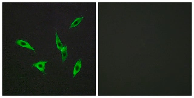
Anti-A1 Adenosine Receptor is specific for A1 adenosine receptor adenosine receptor subunit. By immunoblotting, it reacts strongly with human and rat, and weakly with bovine A1. The antibody detects A1 adenosine receptor in human hippocampus by immunohistology and may be used for immunoprecipitation and confocal microscopy
Immunogen
synthetic peptide (Gln-Pro-Lys-Pro-Pro-Ile-Asp-Glu-Asp-Leu-Pro-Glu-Glu-Lys-Ala-Lys-Ala-Glu-Asp) derived from amino acids 309-326 of the rat A1 adenosine receptor C-terminal domain.
Application
Rabbit anti-A1 adenosine receptor antibody can be used for immunohistochemistry and immunoblotting assays using rat tissues. The antibody can also be used for immunocytochemistry assays using rat spinal cord tissues. The product may also be used for immunoprecipitation applications.
Physical form
Solution in phosphate buffered saline containing 1 mg/mL BSA and 0.05% sodium azide.
Disclaimer
Unless otherwise stated in our catalog or other company documentation accompanying the product(s), our products are intended for research use only and are not to be used for any other purpose, which includes but is not limited to, unauthorized commercial uses, in vitro diagnostic uses, ex vivo or in vivo therapeutic uses or any type of consumption or application to humans or animals.
Not finding the right product?
Try our Product Selector Tool.
Storage Class Code
10 - Combustible liquids
WGK
WGK 1
Flash Point(F)
Not applicable
Flash Point(C)
Not applicable
Personal Protective Equipment
dust mask type N95 (US), Eyeshields, Gloves
Choose from one of the most recent versions:
Already Own This Product?
Find documentation for the products that you have recently purchased in the Document Library.
Stefania Gessi et al.
Advances in pharmacology (San Diego, Calif.), 61, 41-75 (2011-05-19)
The adenosine receptors A(1), A(2A), A(2B), and A(3) are important and ubiquitous mediators of cellular signaling, which play vital roles in protecting tissues and organs from damage. In particular, adenosine triggers tissue protection and repair by different receptor-mediated mechanisms, including
Christopher S Rex et al.
The Journal of neuroscience : the official journal of the Society for Neuroscience, 25(25), 5956-5966 (2005-06-25)
Memory loss in humans begins early in adult life and progresses thereafter. It is not known whether these losses reflect the failure of cellular processes that encode memory or disturbances in events that retrieve it. Here, we report that impairments
Development of an antiserum to rat-brain A1 adenosine receptor: application for immunological and structural comparison of A1 adenosine receptors from various tissues and species.
Nakata, H.
Biochimica et Biophysica Acta, 117, 93-93 (1993)
M Schindler et al.
Neuroscience letters, 297(3), 211-215 (2001-01-04)
Adenosine exerts its physiological actions by binding to G-protein coupled receptors, four of which have been identified and cloned to date (A1, A2a, A2b and A3). Here we report the development of anti-human adenosine A1, receptor anti-peptide polyclonal antibodies and
Role of adenosine A1 and A3 receptors in regulation of cardiomyocyte homeostasis after mitochondrial respiratory chain injury
Schneyvays V et al
American Journal of Physiology. Heart and Circulatory Physiology, 288, H2792-H2792 (2004)
Our team of scientists has experience in all areas of research including Life Science, Material Science, Chemical Synthesis, Chromatography, Analytical and many others.
Contact Technical Service