Skip to Content
Merck
All Photos(1)

Key Documents

MABF1495Z

Sigma-Aldrich

Anti-NK1.1 (mouse) Antibody, clone PK136, Azide Free

clone PK136, from mouse

Synonym(s):

Killer cell lectin-like receptor subfamily B member 1B allele A, Killer cell lectin-like receptor subfamily B member 1C, CD161b, CD161c, CD161 antigen-like family member B, CD161 antigen-like family member C, Ly-55b, Ly-55c, Lymphocyte antigen 55b, Lymph

Sign Into View Organizational & Contract Pricing


About This Item

UNSPSC Code:
12352203
eCl@ss:
32160702
NACRES:
NA.43
clone:
PK136, monoclonal
application:
FACS
IP
activity assay
species reactivity:
mouse
technique(s):
activity assay: suitable
flow cytometry: suitable
immunoprecipitation (IP): suitable
citations:
3

biological source

mouse

Quality Level

antibody form

purified immunoglobulin

antibody product type

primary antibodies

clone

PK136, monoclonal

species reactivity

mouse

technique(s)

activity assay: suitable
flow cytometry: suitable
immunoprecipitation (IP): suitable

isotype

IgG2aκ

GenBank accession no.

NCBI accession no.

UniProt accession no.

target post-translational modification

unmodified

Gene Information

mouse ... Klrb1C(17059)

General description

The NK cell receptor protein 1 (NKR-P1), a.k.a. CD161, molecules represent a family of type II transmembrane C-type lectin-like receptors expressed predominantly by NK cells. A total of five murine NKR-P1-coding genes (Klrb1a/b/c/d/f) have been identified, with NKR-P1A/C/F (CD161a/c/f) functioning as stimulatory receptors that possess a charged transmembrane arginine esidue thought to be important for association with the FcRγ adaptor protein, and NKR-P1B/D as two inhibitory receptors that possess a consensus cytoplasmic ITIM (L/VxYxxL/I/V) motif. The ITIM motif is shown to mediate NKR-P1B recruitment of Src homology 2 (SH2)-containing protein tyrosine phosphatase-1 (SHP-1) in a phosphorylation-dependent manner. All murine NKR-P1 proteins possess a Cys-X-Cys-Pro (CxCP) motif that mediates association with the Src-related nonreceptor protein tyrosine kinase p56lck. Both NKR-P1B and NKR-P1C functionally associate with p56lck, while mutation of the CxCP motif abolishes NKR-P1B/C-mediated signal transduction. Likewise, mutation of ITIM motif abolishes NKR-P1B SHP-1 association as well as NKR-P1B-dependent inhibitory signaling.

Specificity

Clone PK136 recognizes murine NKR-P1B/CD161b (UniProt P27812) and NK1.1/NKR-P1C/CD161c (UniProt P27814), but not CD161a/NKR-P1A (P27811). Upon activation by PK136 binding, NKR-P1C/NK1.1 signals to activate, while NKR-P1B signals to suppress, NK cells for cytotoxic killing of target cells (Carlyle, J.R., et al. (1999). J. Immunol. 162(10):5917-5923). Epitope is present in both spliced isoforms of murine NK1.1/CD161c reported by UniProt (P27814). Clone PK136 does not react with CD161a (by Klrb1a gene) or C57BL/6 mouse strain-derived CDC161b variant (by Klrb1b gene, also known as Klrb1d). There exists a fifth murine CD161 gene Klrb1f, reactivity of this mAb toward this gene product has not been established.

Immunogen

Epitope: Extracellular domain.
Mouse spleen and bone marrow cells (Koo, G.C., and Peppard, J.R. (1984). Hybridoma. 3(3):301-303).

Application

Activity Assay: Representative lots, when administered to mice via i.p. injection, depleted splenic natural killer (NK) cell activity in a dose-dependent manner (Xiao, T.Z., et al. (2012). Biol. Blood Marrow Transplant. 18(2):200-209; Seaman, W.E., et al. (1987). J. Immunol. 138(12):4539-4544).
Activity Assay: A representative lot activated target cell killing by cross-linking NK cell surface NK1.1 (NKR-P1C) and target cell surface Fc receptor. Ab-induced redirected lysis (AIRL) assay employing NK cells expressing both NKR-P1B and NKR-P1C prevented NK cell cytotoxicity activation by clone PK136 (Carlyle, J.R., et al. (1999). J. Immunol. 162(10):5917-5923).
Immunoprecipitation Analysis: A representative lot co-immunoprecipitated SHP-1, but not SHP-2 or SHIP, from Swiss NIH mouse-derived MNK-1 pre-NK cells upon upregulating NKR-P1B intracellular ITIM motif tyrosine phosphorylation by pervanadate treatment (Carlyle, J.R., et al. (1999). J. Immunol. 162(10):5917-5923).
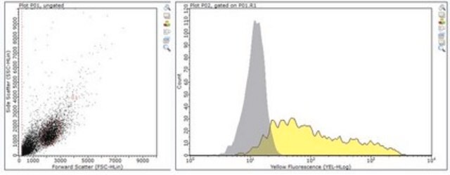
Flow Cytometry Analysis: A representative lot immunostained the surface of Jurkat transfectants expressing C57BL/6J (B6) mouse-derived NKR-P1B/CD161b or Swiss NIH (Sw) mouse-derived NKR-P1C/CD161c/NK1.1, but not Jurkat transfectants expressing B6-derived NKR-P1A/CD161a (Carlyle, J.R., et al. (1999). J. Immunol. 162(10):5917-5923).
Flow Cytometry Analysis: A representative lot immunostained NKR-P1B/CD161b-expressing NK cells from Swiss NIH (Sw) mouse strain, NKR-P1C/CD161c/NK1.1-expressing NK cells from C57BL/6J (B6), as well as (B6 3 Sw)F1 cross-strain-derived NK cells expressing both NKR-P1B and NKR-P1C (Carlyle, J.R., et al. (1999). J. Immunol. 162(10):5917-5923).
Note: Clone PK136 is also available in the following conjugated forms for flow cytometry application, APC (MABF1487 & MABF1488), FITC (MABF1489 & MABF1490), PE (MABF1491), PerCP-Cy5.5 (MABF1493 & MABF1494). redFluor® 710 (MABF1496 & MABF1497).
Anti-NK1.1 (mouse) Antibody, clone PK136, Azide Free is an antibody against NK1.1 (mouse) for use in Flow Cytometry, Activity Assay, Immunohistochemistry, Immunoprecipitation.
Research Category
Inflammation & Immunology
Research Sub Category
Immunological Signaling

Quality

Evaluated by Flow Cytometry in mouse splenocytes.

Flow Cytometry Analysis: 1.0 µg of this antibody detected NK1.1-positive mouse splenocytes.

Target description

25.08 kDa (NK1.1/CD161c isoform 1) and 24.77 kDa (NK1.1/CD161c isoform 2) calculated.

Physical form

Format: Purified
Protein G purified.
Purified mouse monoclonal IgG2aκ antibody in PBS without preservatives.

Storage and Stability

Stable for 1 year at -20°C from date of receipt.
Handling Recommendations: Upon receipt and prior to removing the cap, centrifuge the vial and gently mix the solution. Aliquot into microcentrifuge tubes and store at -20°C. Avoid repeated freeze/thaw cycles, which may damage IgG and affect product performance.

Other Notes

Concentration: Please refer to lot specific datasheet.

Legal Information

GenBank is a registered trademark of United States Department of Health and Human Services
redFluor is a registered trademark of Tonbo Biotechnologies

Disclaimer

Unless otherwise stated in our catalog or other company documentation accompanying the product(s), our products are intended for research use only and are not to be used for any other purpose, which includes but is not limited to, unauthorized commercial uses, in vitro diagnostic uses, ex vivo or in vivo therapeutic uses or any type of consumption or application to humans or animals.

Not finding the right product?  

Try our Product Selector Tool.

Storage Class Code

12 - Non Combustible Liquids

WGK

WGK 2

Flash Point(F)

Not applicable

Flash Point(C)

Not applicable


Certificates of Analysis (COA)

Search for Certificates of Analysis (COA) by entering the products Lot/Batch Number. Lot and Batch Numbers can be found on a product’s label following the words ‘Lot’ or ‘Batch’.

Already Own This Product?

Find documentation for the products that you have recently purchased in the Document Library.

Visit the Document Library

Yao Huang et al.
Molecular oncology, 14(3), 657-668 (2020-01-04)
Oncolytic viruses armed with therapeutic transgenes of interest show great potential in cancer immunotherapy. Here, a novel oncolytic adenovirus carrying a signal regulatory protein-α (SIRPα)-IgG1 Fc fusion gene (termed SG635-SF) was constructed, which could block the CD47 'don't eat me' signal
Jan P Böttcher et al.
Cell, 172(5), 1022-1037 (2018-02-13)
Conventional type 1 dendritic cells (cDC1) are critical for antitumor immunity, and their abundance within tumors is associated with immune-mediated rejection and the success of immunotherapy. Here, we show that cDC1 accumulation in mouse tumors often depends on natural killer
Philipp A Ilinykh et al.
Cell host & microbe, 27(6), 976-991 (2020-04-23)
Marburg virus (MARV) and Ebola virus (EBOV) belong to the family Filoviridae. MARV causes severe disease in humans with high fatality. We previously isolated a large panel of monoclonal antibodies (mAbs) from B cells of a human survivor with previous

Our team of scientists has experience in all areas of research including Life Science, Material Science, Chemical Synthesis, Chromatography, Analytical and many others.

Contact Technical Service